 Movie
Movie Controller


+ Open data

- Basic information
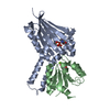
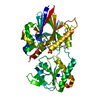
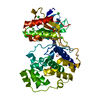
| Entry | Database: PDB / ID: 2dd7 | |||||||||
|---|---|---|---|---|---|---|---|---|---|---|
| Title | A GFP-like protein from marine copepod, Chiridius poppei | |||||||||
 Components Components | green fluorescent protein | |||||||||
 Keywords Keywords | LUMINESCENT PROTEIN / FLUORESCENT PROTEIN | |||||||||
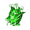
| Function / homology | Green Fluorescent Protein / Green fluorescent protein / Green fluorescent protein-related / Green fluorescent protein / Green fluorescent protein / bioluminescence / Beta Barrel / Mainly Beta / Green fluorescent protein Function and homology information Function and homology information | |||||||||
| Biological species |  Chiridius poppei (arthropod) Chiridius poppei (arthropod) | |||||||||
| Method |  X-RAY DIFFRACTION / SYNCHROTRON / MOLECULAR REPLACEMENT / Resolution: 1.9 Å X-RAY DIFFRACTION / SYNCHROTRON / MOLECULAR REPLACEMENT / Resolution: 1.9 Å | |||||||||
 Authors Authors | Suto, K. / Masuda, H. / Takenaka, Y. / Mizuno, H. | |||||||||
 Citation Citation | Journal: Genes Cells / Year: 2009 Title: Structural basis for red-shifted emission of a GFP-like protein from the marine copepod Chiridius poppei Authors: Suto, K. / Masuda, H. / Takenaka, Y. / Tsuji, F.I. / Mizuno, H. | |||||||||
| History |
|
- Structure visualization
| Structure viewer | Molecule: MolmilJmol/JSmol |
|---|
- Downloads & links
-Download
| PDBx/mmCIF format | 2dd7.cif.gz | 108.1 KB | Display | PDBx/mmCIF format |
|---|---|---|---|---|
| PDB format | pdb2dd7.ent.gz | 83.3 KB | Display | PDB format |
| PDBx/mmJSON format | 2dd7.json.gz | Tree view | PDBx/mmJSON format | |
| Others |  Other downloads Other downloads |
-Validation report
| Arichive directory | https://data.pdbj.org/pub/pdb/validation_reports/dd/2dd7ftp://data.pdbj.org/pub/pdb/validation_reports/dd/2dd7 | HTTPS FTP |
|---|
-Related structure data


-Links
 PDBj
PDBj

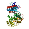
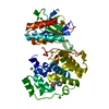
- Assembly
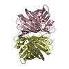
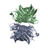
| Deposited unit | 
| |||||||||
|---|---|---|---|---|---|---|---|---|---|---|
| 1 | 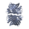
| |||||||||
| 2 | 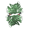
| |||||||||
| Unit cell |
| |||||||||
| Components on special symmetry positions |
|
-Components
| #1: Protein | Mass: 24620.863 Da / Num. of mol.: 2 Source method: isolated from a genetically manipulated source Source: (gene. exp.) Chiridius poppei (arthropod) / Plasmid: pET-101 / Production host:  #2: Chemical | ChemComp-CL / |   Mass: 35.453 Da / Num. of mol.: 1 / Source method: obtained synthetically / Formula: Cl Mass: 35.453 Da / Num. of mol.: 1 / Source method: obtained synthetically / Formula: Cl#3: Chemical | ChemComp-CXS / |   Mass: 221.317 Da / Num. of mol.: 1 / Source method: obtained synthetically / Formula: C9H19NO3S / Comment: pH buffer*YM Mass: 221.317 Da / Num. of mol.: 1 / Source method: obtained synthetically / Formula: C9H19NO3S / Comment: pH buffer*YM#4: Water | ChemComp-HOH / |  Mass: 18.015 Da / Num. of mol.: 436 / Source method: isolated from a natural source / Formula: H2O Mass: 18.015 Da / Num. of mol.: 436 / Source method: isolated from a natural source / Formula: H2OHas protein modification | Y | Sequence details | GLY 55, TYR 56 AND GLY 57 ARE MODIFIED TO MAKE CHROMOPHOR | |
|---|
-Experimental details
-Experiment
| Experiment | Method: X-RAY DIFFRACTION / Number of used crystals: 1 |
|---|
- Sample preparation
| Crystal | Density Matthews: 4.18 Å3/Da / Density % sol: 70.59 % |
|---|---|
| Crystal grow | Temperature: 291 K / Method: vapor diffusion, sitting drop / pH: 10.5 Details: 0.1M CAPS (pH 10.5), 2.2M Ammonium Sulfate, 0.2M NaCl, VAPOR DIFFUSION, SITTING DROP, temperature 291K |
-Data collection
| Diffraction | Mean temperature: 100 K |
|---|---|
| Diffraction source | Source: SYNCHROTRON / Site: Photon Factory  / Beamline: BL-6A / Wavelength: 0.97986 Å / Beamline: BL-6A / Wavelength: 0.97986 Å |
| Detector | Type: ADSC QUANTUM 4 / Detector: CCD / Date: Oct 1, 2004 |
| Radiation | Monochromator: SILICON (1 1 1) / Protocol: SINGLE WAVELENGTH / Monochromatic (M) / Laue (L): M / Scattering type: x-ray |
| Radiation wavelength | Wavelength: 0.97986 Å / Relative weight: 1 |
| Reflection | Resolution: 1.9→30 Å / Num. obs: 65109 / % possible obs: 99.9 % / Observed criterion σ(F): 0 / Observed criterion σ(I): 2 / Biso Wilson estimate: 9.9 Å2 |
| Reflection shell | Resolution: 1.9→2 Å / % possible all: 100 |
- Processing
| Software |
| ||||||||||||||||||||||||||||||||||||||||||||||||||||||||||||||||||||||||||||||||
|---|---|---|---|---|---|---|---|---|---|---|---|---|---|---|---|---|---|---|---|---|---|---|---|---|---|---|---|---|---|---|---|---|---|---|---|---|---|---|---|---|---|---|---|---|---|---|---|---|---|---|---|---|---|---|---|---|---|---|---|---|---|---|---|---|---|---|---|---|---|---|---|---|---|---|---|---|---|---|---|---|---|
| Refinement | Method to determine structure: MOLECULAR REPLACEMENT / Resolution: 1.9→28.82 Å / Rfactor Rfree error: 0.004 / Data cutoff high absF: 3445681.75 / Data cutoff low absF: 0 / Isotropic thermal model: RESTRAINED / Cross valid method: THROUGHOUT / σ(F): 0 / Stereochemistry target values: Engh & Huber
| ||||||||||||||||||||||||||||||||||||||||||||||||||||||||||||||||||||||||||||||||
| Solvent computation | Solvent model: FLAT MODEL / Bsol: 47.9488 Å2 / ksol: 0.393849 e/Å3 | ||||||||||||||||||||||||||||||||||||||||||||||||||||||||||||||||||||||||||||||||
| Displacement parameters | Biso mean: 23.2 Å2
| ||||||||||||||||||||||||||||||||||||||||||||||||||||||||||||||||||||||||||||||||
| Refine analyze |
| ||||||||||||||||||||||||||||||||||||||||||||||||||||||||||||||||||||||||||||||||
| Refinement step | Cycle: LAST / Resolution: 1.9→28.82 Å
| ||||||||||||||||||||||||||||||||||||||||||||||||||||||||||||||||||||||||||||||||
| Refine LS restraints |
| ||||||||||||||||||||||||||||||||||||||||||||||||||||||||||||||||||||||||||||||||
| LS refinement shell | Resolution: 1.9→2.02 Å / Rfactor Rfree error: 0.011 / Total num. of bins used: 6
| ||||||||||||||||||||||||||||||||||||||||||||||||||||||||||||||||||||||||||||||||
| Xplor file |
|
