 Movie
Movie Controller

[English] 日本語
 Yorodumi
- PDB-2d7u: Crystal structure of hypothetical adenylosuccinate synthetase, PH... -
+ Open data

- Basic information
| Entry | Database: PDB / ID: 2d7u | ||||||
|---|---|---|---|---|---|---|---|
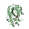
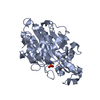
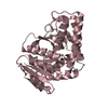
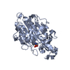
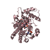
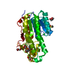
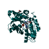
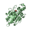
| Title | Crystal structure of hypothetical adenylosuccinate synthetase, PH0438 from Pyrococcus horikoshii OT3 | ||||||
 Components Components | Adenylosuccinate synthetase | ||||||
 Keywords Keywords | LIGASE / Structural Genomics / conserved hypothetical protein / NPPSFA / National Project on Protein Structural and Functional Analyses / RIKEN Structural Genomics/Proteomics Initiative / RSGI | ||||||
| Function / homology |  Function and homology information Function and homology informationadenylosuccinate synthase / adenylosuccinate synthase activity / IMP metabolic process / 'de novo' AMP biosynthetic process / GTP binding / magnesium ion binding / cytoplasm Similarity search - Function | ||||||
| Biological species |   Pyrococcus horikoshii (archaea) Pyrococcus horikoshii (archaea) | ||||||
| Method |  X-RAY DIFFRACTION / SYNCHROTRON / MOLECULAR REPLACEMENT / Resolution: 2.5 Å X-RAY DIFFRACTION / SYNCHROTRON / MOLECULAR REPLACEMENT / Resolution: 2.5 Å | ||||||
 Authors Authors | Xie, Y. / Kishisita, S. / Murayama, K. / Shirouzu, M. / Yokoyama, S. / RIKEN Structural Genomics/Proteomics Initiative (RSGI) | ||||||
 Citation Citation | Journal: To be Published Title: Crystal structure of hypothetical adenylosuccinate synthetase, PH0438 from Pyrococcus horikoshii OT3 Authors: Xie, Y. / Kishishita, S. / Murayama, K. / Shirouzu, M. / Yokoyama, S. | ||||||
| History |
|
- Structure visualization
| Structure viewer | Molecule: MolmilJmol/JSmol |
|---|
- Downloads & links
-Download
| PDBx/mmCIF format | 2d7u.cif.gz | 77.1 KB | Display | PDBx/mmCIF format |
|---|---|---|---|---|
| PDB format | pdb2d7u.ent.gz | 57.3 KB | Display | PDB format |
| PDBx/mmJSON format | 2d7u.json.gz | Tree view | PDBx/mmJSON format | |
| Others |  Other downloads Other downloads |
-Validation report
| Arichive directory | https://data.pdbj.org/pub/pdb/validation_reports/d7/2d7uftp://data.pdbj.org/pub/pdb/validation_reports/d7/2d7u | HTTPS FTP |
|---|
-Related structure data
| Related structure data |  5k7xC 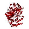 1sooS S: Starting model for refinement C: citing same article ( |
|---|---|
| Similar structure data | |
| Other databases |


-Links
 PDBj
PDBj- Assembly
| Deposited unit | 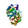
| ||||||||
|---|---|---|---|---|---|---|---|---|---|
| 1 |
| ||||||||
| 2 | 
| ||||||||
| Unit cell |
| ||||||||
| Details | In this crystal packing, it looks like monomer. But, there is no experimental evidence. |
-Components
| #1: Protein | Mass: 37280.395 Da / Num. of mol.: 1 Source method: isolated from a genetically manipulated source Source: (gene. exp.) Pyrococcus horikoshii (archaea) / Strain: OT3 / Plasmid: pET19b / Species (production host): Escherichia coli / Production host:  |
|---|---|
| #2: Water | ChemComp-HOH /  Mass: 18.015 Da / Num. of mol.: 106 / Source method: isolated from a natural source / Formula: H2O Mass: 18.015 Da / Num. of mol.: 106 / Source method: isolated from a natural source / Formula: H2O |
-Experimental details
-Experiment
| Experiment | Method: X-RAY DIFFRACTION / Number of used crystals: 1 |
|---|
- Sample preparation
| Crystal | Density Matthews: 2.17 Å3/Da / Density % sol: 43.23 % |
|---|---|
| Crystal grow | Temperature: 291 K / Method: oil batch / pH: 8.7 Details: 13.5% PEG 20000, 0.1M Tris-HCl, pH 8.7, OIL BATCH, temperature 291K |
-Data collection
| Diffraction | Mean temperature: 100 K |
|---|---|
| Diffraction source | Source: SYNCHROTRON / Site: SPring-8  / Beamline: BL26B2 / Wavelength: 1 Å / Beamline: BL26B2 / Wavelength: 1 Å |
| Detector | Type: RIGAKU JUPITER 210 / Detector: CCD / Date: Aug 27, 2005 / Details: mirrors |
| Radiation | Monochromator: silicon / Protocol: SINGLE WAVELENGTH / Monochromatic (M) / Laue (L): M / Scattering type: x-ray |
| Radiation wavelength | Wavelength: 1 Å / Relative weight: 1 |
| Reflection | Resolution: 2.45→50 Å / Num. obs: 11717 / % possible obs: 96.5 % / Observed criterion σ(I): -3 / Redundancy: 10.2 % / Biso Wilson estimate: 34.9 Å2 / Rsym value: 0.064 / Net I/σ(I): 38.2 |
| Reflection shell | Resolution: 2.45→2.54 Å / Redundancy: 7.3 % / Mean I/σ(I) obs: 6.2 / Num. unique all: 767 / Rsym value: 0.184 / % possible all: 64.6 |
- Processing
| Software |
| ||||||||||||||||||||
|---|---|---|---|---|---|---|---|---|---|---|---|---|---|---|---|---|---|---|---|---|---|
| Refinement | Method to determine structure: MOLECULAR REPLACEMENT Starting model: PDB ENTRY 1SOO Resolution: 2.5→47.58 Å / Rfactor Rfree error: 0.012 / Data cutoff high absF: 770420.1 / Data cutoff low absF: 0 / Isotropic thermal model: RESTRAINED / Cross valid method: THROUGHOUT / σ(F): 0
| ||||||||||||||||||||
| Solvent computation | Solvent model: FLAT MODEL / Bsol: 47.9739 Å2 / ksol: 0.368351 e/Å3 | ||||||||||||||||||||
| Displacement parameters | Biso mean: 42.1 Å2
| ||||||||||||||||||||
| Refine analyze |
| ||||||||||||||||||||
| Refinement step | Cycle: LAST / Resolution: 2.5→47.58 Å
| ||||||||||||||||||||
| Refine LS restraints |
| ||||||||||||||||||||
| LS refinement shell | Resolution: 2.5→2.66 Å / Rfactor Rfree error: 0.042 / Total num. of bins used: 6
| ||||||||||||||||||||
| Xplor file |
|
