 Movie
Movie Controller

[English] 日本語
 Yorodumi
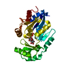
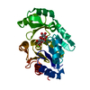
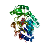
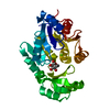
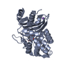
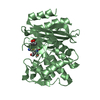
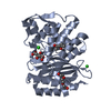
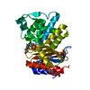
- PDB-2d2v: X-ray structure of the sucrose-phosphatase (SPP) from Synechocyst... -
+ Open data

- Basic information
| Entry | Database: PDB / ID: 2d2v | |||||||||
|---|---|---|---|---|---|---|---|---|---|---|
| Title | X-ray structure of the sucrose-phosphatase (SPP) from Synechocystis sp.PCC6803 in complex with maltose | |||||||||
 Components Components | hypothetical protein slr0953 | |||||||||
 Keywords Keywords | HYDROLASE / phosphohydrolase / HAD superfamily / maltose / cyanobacteria | |||||||||
| Function / homology |  Function and homology information Function and homology informationsucrose-phosphate phosphatase / sucrose-phosphate phosphatase activity / sucrose biosynthetic process / magnesium ion binding Similarity search - Function | |||||||||
| Biological species |  | |||||||||
| Method |  X-RAY DIFFRACTION / SYNCHROTRON / rigid body / Resolution: 2.5 Å X-RAY DIFFRACTION / SYNCHROTRON / rigid body / Resolution: 2.5 Å | |||||||||
 Authors Authors | Fieulaine, S. / Lunn, J.E. / Ferrer, J.-L. | |||||||||
 Citation Citation | Journal: Proteins / Year: 2007 Title: Crystal structure of a cyanobacterial sucrose-phosphatase in complex with glucose-containing disaccharides Authors: Fieulaine, S. / Lunn, J.E. / Ferrer, J.-L. | |||||||||
| History |
|
- Structure visualization
| Structure viewer | Molecule: MolmilJmol/JSmol |
|---|
- Downloads & links
-Download
| PDBx/mmCIF format | 2d2v.cif.gz | 69.6 KB | Display | PDBx/mmCIF format |
|---|---|---|---|---|
| PDB format | pdb2d2v.ent.gz | 49.8 KB | Display | PDB format |
| PDBx/mmJSON format | 2d2v.json.gz | Tree view | PDBx/mmJSON format | |
| Others |  Other downloads Other downloads |
-Validation report
| Arichive directory | https://data.pdbj.org/pub/pdb/validation_reports/d2/2d2vftp://data.pdbj.org/pub/pdb/validation_reports/d2/2d2v | HTTPS FTP |
|---|
-Related structure data
| Related structure data |  2b1qC 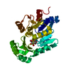 2b1rC 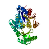 1tj3S C: citing same article ( S: Starting model for refinement |
|---|---|
| Similar structure data |


-Links
 PDBj
PDBj


- Assembly
| Deposited unit | 
| ||||||||
|---|---|---|---|---|---|---|---|---|---|
| 1 |
| ||||||||
| Unit cell |
|
-Components
| #1: Protein | Mass: 27789.867 Da / Num. of mol.: 1 Source method: isolated from a genetically manipulated source Source: (gene. exp.) |
|---|---|
| #2: Polysaccharide | alpha-D-glucopyranose-(1-4)-alpha-D-glucopyranose / alpha-maltose  Source method: isolated from a genetically manipulated source Details: oligosaccharide / References: alpha-maltose |
| #3: Chemical | ChemComp-MG /   Mass: 24.305 Da / Num. of mol.: 1 / Source method: obtained synthetically / Formula: Mg Mass: 24.305 Da / Num. of mol.: 1 / Source method: obtained synthetically / Formula: Mg |
| #4: Water | ChemComp-HOH /  Mass: 18.015 Da / Num. of mol.: 246 / Source method: isolated from a natural source / Formula: H2O Mass: 18.015 Da / Num. of mol.: 246 / Source method: isolated from a natural source / Formula: H2O |
-Experimental details
-Experiment
| Experiment | Method: X-RAY DIFFRACTION / Number of used crystals: 1 |
|---|
- Sample preparation
| Crystal | Density Matthews: 3.31 Å3/Da / Density % sol: 62.8 % |
|---|---|
| Crystal grow | Temperature: 291 K / Method: vapor diffusion, hanging drop / pH: 8 Details: NaFormate, Tris, pH 8.0, VAPOR DIFFUSION, HANGING DROP, temperature 291K |
-Data collection
| Diffraction | Mean temperature: 100 K |
|---|---|
| Diffraction source | Source: SYNCHROTRON / Site: ESRF  / Beamline: ID14-1 / Wavelength: 0.934 Å / Beamline: ID14-1 / Wavelength: 0.934 Å |
| Detector | Type: ADSC QUANTUM 4 / Detector: CCD / Date: Nov 26, 2004 |
| Radiation | Monochromator: Diamond(111), Ge(220) / Protocol: SINGLE WAVELENGTH / Monochromatic (M) / Laue (L): M / Scattering type: x-ray |
| Radiation wavelength | Wavelength: 0.934 Å / Relative weight: 1 |
| Reflection | Resolution: 2.5→50 Å / Num. obs: 13929 / % possible obs: 99.9 % / Observed criterion σ(F): 2 / Observed criterion σ(I): 2 / Rmerge(I) obs: 0.17 / Rsym value: 0.17 / Net I/σ(I): 13.5 |
- Processing
| Software |
| |||||||||||||||||||||||||
|---|---|---|---|---|---|---|---|---|---|---|---|---|---|---|---|---|---|---|---|---|---|---|---|---|---|---|
| Refinement | Method to determine structure: rigid body Starting model: 1tj3 Resolution: 2.5→50 Å / σ(F): 2 / σ(I): 2 / Stereochemistry target values: Engh & Huber
| |||||||||||||||||||||||||
| Refinement step | Cycle: LAST / Resolution: 2.5→50 Å
| |||||||||||||||||||||||||
| Refine LS restraints |
|
