 Movie
Movie Controller

[English] 日本語
 Yorodumi
- PDB-2ciq: Structure-based functional annotation: Yeast ymr099c codes for a ... -
+ Open data

- Basic information
| Entry | Database: PDB / ID: 2ciq | ||||||
|---|---|---|---|---|---|---|---|
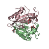
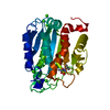
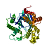
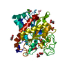
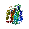
| Title | Structure-based functional annotation: Yeast ymr099c codes for a D- hexose-6-phosphate mutarotase. | ||||||
 Components Components | HEXOSE-6-PHOSPHATE MUTAROTASE | ||||||
 Keywords Keywords | ISOMERASE / EPIMERASE / MANNOSE-6-PHOSPHATE-1-EPIMERASE / GALACTOSE-6-PHOSPHATE-1-EPIMERASE / D-HEXOSE-6-PHOSPHATE-1-MUTAROTASE | ||||||
| Function / homology |  Function and homology information Function and homology informationglucose-6-phosphate 1-epimerase / glucose-6-phosphate 1-epimerase activity / carbohydrate binding / carbohydrate metabolic process / nucleus / cytoplasm Similarity search - Function | ||||||
| Biological species |  | ||||||
| Method |  X-RAY DIFFRACTION / SYNCHROTRON / MOLECULAR REPLACEMENT / Resolution: 1.7 Å X-RAY DIFFRACTION / SYNCHROTRON / MOLECULAR REPLACEMENT / Resolution: 1.7 Å | ||||||
 Authors Authors | Graille, M. / Baltaze, J.-P. / Leulliot, N. / Liger, D. / Quevillon-Cheruel, S. / van Tilbeurgh, H. | ||||||
 Citation Citation | Journal: J. Biol. Chem. / Year: 2006 Title: Structure-based functional annotation: yeast ymr099c codes for a D-hexose-6-phosphate mutarotase. Authors: Graille, M. / Baltaze, J.P. / Leulliot, N. / Liger, D. / Quevillon-Cheruel, S. / van Tilbeurgh, H. | ||||||
| History |
| ||||||
| Remark 700 | SHEET THE SHEET STRUCTURE OF THIS MOLECULE IS BIFURCATED. IN ORDER TO REPRESENT THIS FEATURE IN ... SHEET THE SHEET STRUCTURE OF THIS MOLECULE IS BIFURCATED. IN ORDER TO REPRESENT THIS FEATURE IN THE SHEET RECORDS BELOW, TWO SHEETS ARE DEFINED. |
- Structure visualization
| Structure viewer | Molecule: MolmilJmol/JSmol |
|---|
- Downloads & links
-Download
| PDBx/mmCIF format | 2ciq.cif.gz | 81.4 KB | Display | PDBx/mmCIF format |
|---|---|---|---|---|
| PDB format | pdb2ciq.ent.gz | 58.3 KB | Display | PDB format |
| PDBx/mmJSON format | 2ciq.json.gz | Tree view | PDBx/mmJSON format | |
| Others |  Other downloads Other downloads |
-Validation report
| Arichive directory | https://data.pdbj.org/pub/pdb/validation_reports/ci/2ciqftp://data.pdbj.org/pub/pdb/validation_reports/ci/2ciq | HTTPS FTP |
|---|
-Related structure data
| Related structure data | 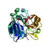 2cirC  2cisC 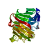 1jovS C: citing same article ( S: Starting model for refinement |
|---|---|
| Similar structure data |


-Links
 PDBj
PDBj


- Assembly
| Deposited unit | 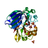
| ||||||||
|---|---|---|---|---|---|---|---|---|---|
| 1 |
| ||||||||
| Unit cell |
|
-Components
-Protein , 1 types, 1 molecules A
| #1: Protein | Mass: 33995.773 Da / Num. of mol.: 1 Source method: isolated from a genetically manipulated source Source: (gene. exp.) Strain: S288C / Plasmid: PET9 / Production host:  References: UniProt: Q03161, glucose-6-phosphate 1-epimerase |
|---|
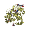
-Non-polymers , 5 types, 308 molecules 


| #2: Chemical | ChemComp-EPE /  Mass: 238.305 Da / Num. of mol.: 1 / Source method: obtained synthetically / Formula: C8H18N2O4S / Comment: pH buffer*YM Mass: 238.305 Da / Num. of mol.: 1 / Source method: obtained synthetically / Formula: C8H18N2O4S / Comment: pH buffer*YM | ||
|---|---|---|---|
| #3: Chemical | ChemComp-NA /  Mass: 22.990 Da / Num. of mol.: 1 / Source method: obtained synthetically / Formula: Na Mass: 22.990 Da / Num. of mol.: 1 / Source method: obtained synthetically / Formula: Na | ||
| #4: Chemical | ChemComp-GOL /  Mass: 92.094 Da / Num. of mol.: 1 / Source method: obtained synthetically / Formula: C3H8O3 Mass: 92.094 Da / Num. of mol.: 1 / Source method: obtained synthetically / Formula: C3H8O3 | ||
| #5: Chemical |  Mass: 96.063 Da / Num. of mol.: 2 / Source method: obtained synthetically / Formula: SO4 Mass: 96.063 Da / Num. of mol.: 2 / Source method: obtained synthetically / Formula: SO4#6: Water | ChemComp-HOH / | Mass: 18.015 Da / Num. of mol.: 303 / Source method: isolated from a natural source / Formula: H2O |
-Experimental details
-Experiment
| Experiment | Method: X-RAY DIFFRACTION / Number of used crystals: 1 |
|---|
- Sample preparation
| Crystal | Density Matthews: 2.8 Å3/Da / Density % sol: 50 % |
|---|---|
| Crystal grow | pH: 7.5 Details: 25-30% POLYETHYLENE GLYCOL 3000, 0.2 M LITHIUM SULFATE, 0.1M HEPES PH 7.5 |
-Data collection
| Diffraction | Mean temperature: 100 K |
|---|---|
| Diffraction source | Source: SYNCHROTRON / Site: ESRF  / Beamline: ID14-1 / Wavelength: 0.978 / Beamline: ID14-1 / Wavelength: 0.978 |
| Detector | Type: ADSC CCD / Detector: CCD / Date: Apr 20, 2005 |
| Radiation | Protocol: SINGLE WAVELENGTH / Monochromatic (M) / Laue (L): M / Scattering type: x-ray |
| Radiation wavelength | Wavelength: 0.978 Å / Relative weight: 1 |
| Reflection | Resolution: 1.7→20 Å / Num. obs: 39082 / % possible obs: 98.4 % / Observed criterion σ(I): 2 / Redundancy: 3.5 % / Rmerge(I) obs: 0.06 / Net I/σ(I): 14.8 |
| Reflection shell | Resolution: 1.7→1.8 Å / Rmerge(I) obs: 0.41 / Mean I/σ(I) obs: 4.9 / % possible all: 98.2 |
- Processing
| Software |
| ||||||||||||||||||||||||||||||||||||||||||||||||||||||||||||||||||||||||||||||||||||||||||||||||||||||||||||||||||||||||||||||||||||||||||||||||||||||||||||||||||||||||||||||||||||||
|---|---|---|---|---|---|---|---|---|---|---|---|---|---|---|---|---|---|---|---|---|---|---|---|---|---|---|---|---|---|---|---|---|---|---|---|---|---|---|---|---|---|---|---|---|---|---|---|---|---|---|---|---|---|---|---|---|---|---|---|---|---|---|---|---|---|---|---|---|---|---|---|---|---|---|---|---|---|---|---|---|---|---|---|---|---|---|---|---|---|---|---|---|---|---|---|---|---|---|---|---|---|---|---|---|---|---|---|---|---|---|---|---|---|---|---|---|---|---|---|---|---|---|---|---|---|---|---|---|---|---|---|---|---|---|---|---|---|---|---|---|---|---|---|---|---|---|---|---|---|---|---|---|---|---|---|---|---|---|---|---|---|---|---|---|---|---|---|---|---|---|---|---|---|---|---|---|---|---|---|---|---|---|---|
| Refinement | Method to determine structure: MOLECULAR REPLACEMENT Starting model: PDB ENTRY 1JOV Resolution: 1.7→19.16 Å / Cor.coef. Fo:Fc: 0.96 / Cor.coef. Fo:Fc free: 0.941 / SU B: 2.072 / SU ML: 0.069 / Cross valid method: THROUGHOUT / ESU R: 0.102 / ESU R Free: 0.101 / Stereochemistry target values: MAXIMUM LIKELIHOOD / Details: HYDROGENS HAVE BEEN ADDED IN THE RIDING POSITIONS.
| ||||||||||||||||||||||||||||||||||||||||||||||||||||||||||||||||||||||||||||||||||||||||||||||||||||||||||||||||||||||||||||||||||||||||||||||||||||||||||||||||||||||||||||||||||||||
| Solvent computation | Ion probe radii: 0.8 Å / Shrinkage radii: 0.8 Å / VDW probe radii: 1.2 Å / Solvent model: MASK | ||||||||||||||||||||||||||||||||||||||||||||||||||||||||||||||||||||||||||||||||||||||||||||||||||||||||||||||||||||||||||||||||||||||||||||||||||||||||||||||||||||||||||||||||||||||
| Displacement parameters | Biso mean: 24.58 Å2
| ||||||||||||||||||||||||||||||||||||||||||||||||||||||||||||||||||||||||||||||||||||||||||||||||||||||||||||||||||||||||||||||||||||||||||||||||||||||||||||||||||||||||||||||||||||||
| Refinement step | Cycle: LAST / Resolution: 1.7→19.16 Å
| ||||||||||||||||||||||||||||||||||||||||||||||||||||||||||||||||||||||||||||||||||||||||||||||||||||||||||||||||||||||||||||||||||||||||||||||||||||||||||||||||||||||||||||||||||||||
| Refine LS restraints |
|