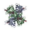
SHEET DETERMINATION METHOD: DSSP THE SHEETS PRESENTED AS "AA" IN EACH CHAIN ON SHEET RECORDS BELOW ... SHEET DETERMINATION METHOD: DSSP THE SHEETS PRESENTED AS "AA" IN EACH CHAIN ON SHEET RECORDS BELOW IS ACTUALLY AN 9-STRANDED BARREL THIS IS REPRESENTED BY A 10-STRANDED SHEET IN WHICH THE FIRST AND LAST STRANDS ARE IDENTICAL. THE SHEETS PRESENTED AS "BA" IN EACH CHAIN ON SHEET RECORDS BELOW IS ACTUALLY AN 9-STRANDED BARREL THIS IS REPRESENTED BY A 10-STRANDED SHEET IN WHICH THE FIRST AND LAST STRANDS ARE IDENTICAL.
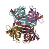
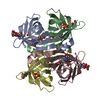
Mass: 14019.936 Da / Num. of mol.: 2 Source method: isolated from a genetically manipulated source Source: (gene. exp.) GALLUS GALLUS (chicken) / Production host: ESCHERICHIA COLI (E. coli)
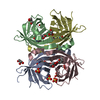
Resolution: 1.75→25 Å / Cor.coef. Fo:Fc: 0.955 / Cor.coef. Fo:Fc free: 0.933 / SU B: 2.731 / SU ML: 0.087 / Cross valid method: THROUGHOUT / ESU R: 0.127 / ESU R Free: 0.123 / Stereochemistry target values: MAXIMUM LIKELIHOOD / Details: HYDROGENS HAVE BEEN ADDED IN THE RIDING POSITIONS.
Rfactor
Num. reflection
% reflection
Selection details
Rfree
0.233
1342
5.1 %
RANDOM
Rwork
0.193
-
-
-
obs
0.195
25104
99.9 %
-
Solvent computation
Ion probe radii: 0.8 Å / Shrinkage radii: 0.8 Å / VDW probe radii: 1.2 Å / Solvent model: MASK
 Movie
Movie Controller


 Open data
 Basic information
 Components
 Keywords
 Function and homology information
 X-RAY DIFFRACTION /
  Authors
 Citation
 Structure visualization
 Downloads & links
 Other downloads
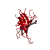


 PDBj
PDBj Assembly


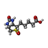
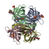
 Mass: 260.310 Da / Num. of mol.: 2 / Source method: obtained synthetically / Formula: C10H16N2O4S
 Mass: 18.015 Da / Num. of mol.: 212 / Source method: isolated from a natural source / Formula: H2O
 Sample preparation
 / Beamline: I711 / Wavelength: 1.063
  Processing