 Movie
Movie Controller

[English] 日本語
 Yorodumi
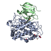
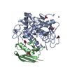
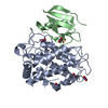
- PDB-2ahk: Crystal structure of the met-form of the copper-bound Streptomyce... -
+ Open data

- Basic information
| Entry | Database: PDB / ID: 2ahk | ||||||
|---|---|---|---|---|---|---|---|
| Title | Crystal structure of the met-form of the copper-bound Streptomyces castaneoglobisporus tyrosinase in complex with a caddie protein obtained by soking in cupric sulfate for 6 months | ||||||
 Components Components |
| ||||||
 Keywords Keywords | OXIDOREDUCTASE/METAL TRANSPORT / TYROSINASE / BINARY COMPLEX / COPPER / OXIDOREDUCTASE-METAL TRANSPORT COMPLEX | ||||||
| Function / homology |  Function and homology information Function and homology informationmelanin biosynthetic process / oxidoreductase activity / copper ion binding / metal ion binding Similarity search - Function | ||||||
| Biological species |  Streptomyces castaneoglobisporus (bacteria) Streptomyces castaneoglobisporus (bacteria) | ||||||
| Method |  X-RAY DIFFRACTION / MOLECULAR REPLACEMENT / Resolution: 1.71 Å X-RAY DIFFRACTION / MOLECULAR REPLACEMENT / Resolution: 1.71 Å | ||||||
 Authors Authors | Matoba, Y. / Kumagai, T. / Yamamoto, A. / Yoshitsu, H. / Sugiyama, M. | ||||||
 Citation Citation | Journal: J.Biol.Chem. / Year: 2006 Title: Crystallographic Evidence That the Dinuclear Copper Center of Tyrosinase Is Flexible during Catalysis Authors: Matoba, Y. / Kumagai, T. / Yamamoto, A. / Yoshitsu, H. / Sugiyama, M. | ||||||
| History |
|
- Structure visualization
| Structure viewer | Molecule: MolmilJmol/JSmol |
|---|
- Downloads & links
-Download
| PDBx/mmCIF format | 2ahk.cif.gz | 92 KB | Display | PDBx/mmCIF format |
|---|---|---|---|---|
| PDB format | pdb2ahk.ent.gz | 67.6 KB | Display | PDB format |
| PDBx/mmJSON format | 2ahk.json.gz | Tree view | PDBx/mmJSON format | |
| Others |  Other downloads Other downloads |
-Validation report
| Arichive directory | https://data.pdbj.org/pub/pdb/validation_reports/ah/2ahkftp://data.pdbj.org/pub/pdb/validation_reports/ah/2ahk | HTTPS FTP |
|---|
-Related structure data
| Related structure data | 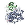 1wx2C  1wx4C  1wx5C  1wxcSC  2ahlC  2zmxC C: citing same article ( S: Starting model for refinement |
|---|---|
| Similar structure data |


-Links
 PDBj
PDBj

- Assembly
| Deposited unit | 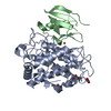
| ||||||||||||
|---|---|---|---|---|---|---|---|---|---|---|---|---|---|
| 1 |
| ||||||||||||
| Unit cell |
| ||||||||||||
| Components on special symmetry positions |
|
-Components
| #1: Protein | Mass: 32089.564 Da / Num. of mol.: 1 Source method: isolated from a genetically manipulated source Source: (gene. exp.) Streptomyces castaneoglobisporus (bacteria)Gene: TYRC / Plasmid: PET-MEL2 / Production host: | ||||
|---|---|---|---|---|---|
| #2: Protein | Mass: 14203.812 Da / Num. of mol.: 1 Source method: isolated from a genetically manipulated source Source: (gene. exp.) Streptomyces castaneoglobisporus (bacteria)Gene: ORF378 / Plasmid: PET-MEL2 / Production host: | ||||
| #3: Chemical |   Mass: 63.546 Da / Num. of mol.: 3 / Source method: obtained synthetically / Formula: Cu Mass: 63.546 Da / Num. of mol.: 3 / Source method: obtained synthetically / Formula: Cu#4: Chemical | ChemComp-NO3 /   Mass: 62.005 Da / Num. of mol.: 4 / Source method: obtained synthetically / Formula: NO3 Mass: 62.005 Da / Num. of mol.: 4 / Source method: obtained synthetically / Formula: NO3#5: Water | ChemComp-HOH / |  Mass: 18.015 Da / Num. of mol.: 315 / Source method: isolated from a natural source / Formula: H2O Mass: 18.015 Da / Num. of mol.: 315 / Source method: isolated from a natural source / Formula: H2O |
-Experimental details
-Experiment
| Experiment | Method: X-RAY DIFFRACTION / Number of used crystals: 1 |
|---|
- Sample preparation
| Crystal | Density Matthews: 1.89 Å3/Da / Density % sol: 34.9 % |
|---|---|
| Crystal grow | Temperature: 298 K / Method: vapor diffusion, sitting drop / pH: 6.5 Details: PEG3350, Sodium nitrate, Hepes, pH 6.5, VAPOR DIFFUSION, SITTING DROP, temperature 298K |
-Data collection
| Diffraction | Mean temperature: 100 K |
|---|---|
| Diffraction source | Source: ROTATING ANODE / Type: RIGAKU / Wavelength: 1.5418 Å |
| Detector | Type: RIGAKU / Detector: IMAGE PLATE / Date: May 24, 2005 |
| Radiation | Protocol: SINGLE WAVELENGTH / Monochromatic (M) / Laue (L): M / Scattering type: x-ray |
| Radiation wavelength | Wavelength: 1.5418 Å / Relative weight: 1 |
| Reflection | Resolution: 1.71→100 Å / Num. all: 37596 / Num. obs: 37596 / % possible obs: 97.7 % / Observed criterion σ(F): 1 / Observed criterion σ(I): 1 / Redundancy: 3.26 % / Biso Wilson estimate: 27 Å2 / Rmerge(I) obs: 0.037 / Rsym value: 0.037 / Net I/σ(I): 15.3 |
| Reflection shell | Resolution: 1.71→1.77 Å / Redundancy: 3.3 % / Rmerge(I) obs: 0.308 / Mean I/σ(I) obs: 3.7 / Num. unique all: 3753 / Rsym value: 0.308 / % possible all: 98.4 |
- Processing
| Software |
| ||||||||||||||||||||||||||||||||||||||||||||||||||||||||||||||||||||||||||||||||
|---|---|---|---|---|---|---|---|---|---|---|---|---|---|---|---|---|---|---|---|---|---|---|---|---|---|---|---|---|---|---|---|---|---|---|---|---|---|---|---|---|---|---|---|---|---|---|---|---|---|---|---|---|---|---|---|---|---|---|---|---|---|---|---|---|---|---|---|---|---|---|---|---|---|---|---|---|---|---|---|---|---|
| Refinement | Method to determine structure: MOLECULAR REPLACEMENT Starting model: PDB ENTRY 1WXC Resolution: 1.71→30 Å / Rfactor Rfree error: 0.006 / Data cutoff high absF: 10000000 / Data cutoff low absF: 0.001 / Isotropic thermal model: RESTRAINED / Cross valid method: THROUGHOUT / σ(F): 2 / σ(I): 1 / Stereochemistry target values: Engh & Huber / Details: BULK SOLVENT MODEL USED
| ||||||||||||||||||||||||||||||||||||||||||||||||||||||||||||||||||||||||||||||||
| Displacement parameters | Biso mean: 25.3 Å2
| ||||||||||||||||||||||||||||||||||||||||||||||||||||||||||||||||||||||||||||||||
| Refine analyze |
| ||||||||||||||||||||||||||||||||||||||||||||||||||||||||||||||||||||||||||||||||
| Refinement step | Cycle: LAST / Resolution: 1.71→30 Å
| ||||||||||||||||||||||||||||||||||||||||||||||||||||||||||||||||||||||||||||||||
| Refine LS restraints |
| ||||||||||||||||||||||||||||||||||||||||||||||||||||||||||||||||||||||||||||||||
| LS refinement shell | Resolution: 1.71→1.82 Å / Rfactor Rfree error: 0.02 / Total num. of bins used: 6
| ||||||||||||||||||||||||||||||||||||||||||||||||||||||||||||||||||||||||||||||||
| Xplor file |
|
