 Movie
Movie Controller

[English] 日本語
 Yorodumi
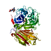
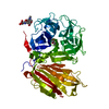
- PDB-2aez: Crystal structure of fructan 1-exohydrolase IIa (E201Q) from Cich... -
+ Open data

- Basic information
| Entry | Database: PDB / ID: 2aez | |||||||||
|---|---|---|---|---|---|---|---|---|---|---|
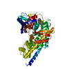
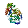
| Title | Crystal structure of fructan 1-exohydrolase IIa (E201Q) from Cichorium intybus in complex with 1-kestose | |||||||||
 Components Components | fructan 1-exohydrolase IIa | |||||||||
 Keywords Keywords | HYDROLASE / five fold beta propeller | |||||||||
| Function / homology |  Function and homology information Function and homology informationfructan beta-fructosidase / fructan beta-fructosidase activity / carbohydrate metabolic process Similarity search - Function | |||||||||
| Biological species |  Cichorium intybus (chicory) Cichorium intybus (chicory) | |||||||||
| Method |  X-RAY DIFFRACTION / SYNCHROTRON / MOLECULAR REPLACEMENT / Resolution: 3.05 Å X-RAY DIFFRACTION / SYNCHROTRON / MOLECULAR REPLACEMENT / Resolution: 3.05 Å | |||||||||
 Authors Authors | Verhaest, M. / Lammens, W. / Le Roy, K. / De Ranter, C.J. / Van Laere, A. / Van den Ende, W. / Rabijns, A. | |||||||||
 Citation Citation | Journal: New Phytol / Year: 2007 Title: Insights into the fine architecture of the active site of chicory fructan 1-exohydrolase: 1-kestose as substrate vs sucrose as inhibitor. Authors: Verhaest, M. / Lammens, W. / Le Roy, K. / De Ranter, C.J. / Van Laere, A. / Rabijns, A. / Van den Ende, W. | |||||||||
| History |
|
- Structure visualization
| Structure viewer | Molecule: MolmilJmol/JSmol |
|---|
- Downloads & links
-Download
| PDBx/mmCIF format | 2aez.cif.gz | 125 KB | Display | PDBx/mmCIF format |
|---|---|---|---|---|
| PDB format | pdb2aez.ent.gz | 95.7 KB | Display | PDB format |
| PDBx/mmJSON format | 2aez.json.gz | Tree view | PDBx/mmJSON format | |
| Others |  Other downloads Other downloads |
-Validation report
| Arichive directory | https://data.pdbj.org/pub/pdb/validation_reports/ae/2aezftp://data.pdbj.org/pub/pdb/validation_reports/ae/2aez | HTTPS FTP |
|---|
-Related structure data
| Related structure data |  2addC  2adeC  2aeyC  1st8S C: citing same article ( S: Starting model for refinement |
|---|---|
| Similar structure data |


-Links
 PDBj
PDBj- Assembly
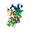
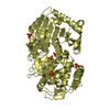
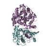
| Deposited unit | 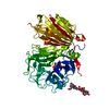
| ||||||||
|---|---|---|---|---|---|---|---|---|---|
| 1 |
| ||||||||
| Unit cell |
|
-Components
| #1: Protein | Mass: 61114.980 Da / Num. of mol.: 1 / Mutation: E201Q Source method: isolated from a genetically manipulated source Source: (gene. exp.) Cichorium intybus (chicory) / Gene: 1-FEH IIa / Production host:  Pichia pastoris (fungus) / References: UniProt: Q93X60, EC: 3.2.1.153 Pichia pastoris (fungus) / References: UniProt: Q93X60, EC: 3.2.1.153 |
|---|---|
| #2: Polysaccharide | 2-acetamido-2-deoxy-alpha-D-glucopyranose-(1-4)-2-acetamido-2-deoxy-beta-D-glucopyranose Source method: isolated from a genetically manipulated source |
| #3: Polysaccharide | beta-D-mannopyranose-(1-3)-beta-D-mannopyranose-(1-4)-2-acetamido-2-deoxy-beta-D-glucopyranose-(1-4) ...beta-D-mannopyranose-(1-3)-beta-D-mannopyranose-(1-4)-2-acetamido-2-deoxy-beta-D-glucopyranose-(1-4)-2-acetamido-2-deoxy-beta-D-glucopyranose Source method: isolated from a genetically manipulated source |
| #4: Polysaccharide | beta-D-fructofuranose-(2-1)-beta-D-fructofuranose-(2-1)-alpha-D-glucopyranose / 1-kestose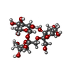  Source method: isolated from a genetically manipulated source Details: oligosaccharide with reducing-end-to-reducing-end glycosidic bond References: 1-kestose |
| #5: Water | ChemComp-HOH /  Mass: 18.015 Da / Num. of mol.: 85 / Source method: isolated from a natural source / Formula: H2O Mass: 18.015 Da / Num. of mol.: 85 / Source method: isolated from a natural source / Formula: H2O |
| Has protein modification | Y |
-Experimental details
-Experiment
| Experiment | Method: X-RAY DIFFRACTION / Number of used crystals: 1 |
|---|
- Sample preparation
| Crystal | Density Matthews: 7.2 Å3/Da / Density % sol: 82 % |
|---|---|
| Crystal grow | Temperature: 277 K / Method: vapor diffusion, hanging drop / pH: 7 Details: sodium potassium phosphate, potassium phosphate, pH 7.0, VAPOR DIFFUSION, HANGING DROP, temperature 277K |
-Data collection
| Diffraction | Mean temperature: 100 K |
|---|---|
| Diffraction source | Source: SYNCHROTRON / Site: EMBL/DESY, HAMBURG  / Beamline: BW7B / Wavelength: 0.8424 Å / Beamline: BW7B / Wavelength: 0.8424 Å |
| Detector | Type: MARRESEARCH / Detector: CCD / Date: May 2, 2005 / Details: bent mirror |
| Radiation | Monochromator: triangular monochromator / Protocol: SINGLE WAVELENGTH / Monochromatic (M) / Laue (L): M / Scattering type: x-ray |
| Radiation wavelength | Wavelength: 0.8424 Å / Relative weight: 1 |
| Reflection | Resolution: 3.05→40 Å / Num. all: 34494 / Num. obs: 27790 / % possible obs: 80.4 % / Observed criterion σ(F): 1.41 / Observed criterion σ(I): 2 / Redundancy: 5.6 % / Rmerge(I) obs: 0.11 |
| Reflection shell | Resolution: 3.05→3.1 Å / Rmerge(I) obs: 0.498 / % possible all: 99.6 |
- Processing
| Software |
| ||||||||||||||||||||||||||||||||||||||||||||||||||||||||||||||||||||||||||||||||
|---|---|---|---|---|---|---|---|---|---|---|---|---|---|---|---|---|---|---|---|---|---|---|---|---|---|---|---|---|---|---|---|---|---|---|---|---|---|---|---|---|---|---|---|---|---|---|---|---|---|---|---|---|---|---|---|---|---|---|---|---|---|---|---|---|---|---|---|---|---|---|---|---|---|---|---|---|---|---|---|---|---|
| Refinement | Method to determine structure: MOLECULAR REPLACEMENT Starting model: PDB ENTRY 1ST8 Resolution: 3.05→29.44 Å / Rfactor Rfree error: 0.003 / Data cutoff high absF: 174791.28 / Data cutoff low absF: 0 / Isotropic thermal model: RESTRAINED / Cross valid method: THROUGHOUT / σ(F): 0 / Stereochemistry target values: Engh & Huber / Details: FRIEDEL PAIRS WERE USED IN REFINEMENT.
| ||||||||||||||||||||||||||||||||||||||||||||||||||||||||||||||||||||||||||||||||
| Solvent computation | Solvent model: FLAT MODEL / Bsol: 58.4799 Å2 / ksol: 0.46991 e/Å3 | ||||||||||||||||||||||||||||||||||||||||||||||||||||||||||||||||||||||||||||||||
| Displacement parameters | Biso mean: 45.4 Å2
| ||||||||||||||||||||||||||||||||||||||||||||||||||||||||||||||||||||||||||||||||
| Refine analyze |
| ||||||||||||||||||||||||||||||||||||||||||||||||||||||||||||||||||||||||||||||||
| Refinement step | Cycle: LAST / Resolution: 3.05→29.44 Å
| ||||||||||||||||||||||||||||||||||||||||||||||||||||||||||||||||||||||||||||||||
| Refine LS restraints |
| ||||||||||||||||||||||||||||||||||||||||||||||||||||||||||||||||||||||||||||||||
| LS refinement shell | Resolution: 3.05→3.24 Å / Rfactor Rfree error: 0.011 / Total num. of bins used: 6
| ||||||||||||||||||||||||||||||||||||||||||||||||||||||||||||||||||||||||||||||||
| Xplor file |
|
