- PDB-2ac5: Structure of human Mnk2 Kinase Domain mutant D228G -
+
Open data
ID or keywords:
Loading...
-
Basic information
Entry
Database: PDB / ID: 2ac5
Title
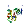
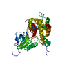
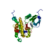
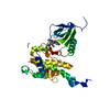
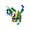
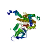
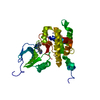
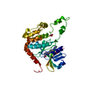
Structure of human Mnk2 Kinase Domain mutant D228G
Components
MAP kinase-interacting serine/threonine kinase 2
Keywords
TRANSFERASE / DFG motif
Function / homology
Function and homology information
calcium-dependent protein serine/threonine kinase activity / cellular response to arsenic-containing substance / calcium/calmodulin-dependent protein kinase activity / hemopoiesis / extrinsic apoptotic signaling pathway in absence of ligand / PML body / regulation of translation / protein phosphorylation / calmodulin binding / non-specific serine/threonine protein kinase ...calcium-dependent protein serine/threonine kinase activity / cellular response to arsenic-containing substance / calcium/calmodulin-dependent protein kinase activity / hemopoiesis / extrinsic apoptotic signaling pathway in absence of ligand / PML body / regulation of translation / protein phosphorylation / calmodulin binding / non-specific serine/threonine protein kinase / cell surface receptor signaling pathway / intracellular signal transduction / nuclear body / protein serine kinase activity / protein serine/threonine kinase activity / nucleoplasm / ATP binding / metal ion binding / nucleus / cytoplasm Similarity search - Function
: / Phosphorylase Kinase; domain 1 / Phosphorylase Kinase; domain 1 / Transferase(Phosphotransferase) domain 1 / Transferase(Phosphotransferase); domain 1 / Serine/threonine-protein kinase, active site / Serine/Threonine protein kinases active-site signature. / Protein kinase domain / Serine/Threonine protein kinases, catalytic domain / Protein kinase, ATP binding site ...: / Phosphorylase Kinase; domain 1 / Phosphorylase Kinase; domain 1 / Transferase(Phosphotransferase) domain 1 / Transferase(Phosphotransferase); domain 1 / Serine/threonine-protein kinase, active site / Serine/Threonine protein kinases active-site signature. / Protein kinase domain / Serine/Threonine protein kinases, catalytic domain / Protein kinase, ATP binding site / Protein kinases ATP-binding region signature. / Protein kinase domain profile. / Protein kinase domain / Protein kinase-like domain superfamily / 2-Layer Sandwich / Orthogonal Bundle / Mainly Alpha / Alpha Beta Similarity search - Domain/homology
In the structure databanks used in Yorodumi, some data are registered as the other names, "COVID-19 virus" and "2019-nCoV". Here are the details of the virus and the list of structure data.
Jan 31, 2019. EMDB accession codes are about to change! (news from PDBe EMDB page)
EMDB accession codes are about to change! (news from PDBe EMDB page)
The allocation of 4 digits for EMDB accession codes will soon come to an end. Whilst these codes will remain in use, new EMDB accession codes will include an additional digit and will expand incrementally as the available range of codes is exhausted. The current 4-digit format prefixed with “EMD-” (i.e. EMD-XXXX) will advance to a 5-digit format (i.e. EMD-XXXXX), and so on. It is currently estimated that the 4-digit codes will be depleted around Spring 2019, at which point the 5-digit format will come into force.
The EM Navigator/Yorodumi systems omit the EMD- prefix.
Related info.:Q: What is EMD? / ID/Accession-code notation in Yorodumi/EM Navigator
Yorodumi is a browser for structure data from EMDB, PDB, SASBDB, etc.
This page is also the successor to EM Navigator detail page, and also detail information page/front-end page for Omokage search.
The word "yorodu" (or yorozu) is an old Japanese word meaning "ten thousand". "mi" (miru) is to see.
Related info.:EMDB / PDB / SASBDB / Comparison of 3 databanks / Yorodumi Search / Aug 31, 2016. New EM Navigator & Yorodumi / Yorodumi Papers / Jmol/JSmol / Function and homology information / Changes in new EM Navigator and Yorodumi
 Movie
Movie Controller


 Open data
 Basic information
 Components
 Keywords
 Function and homology information
 Homo sapiens (human)
 X-RAY DIFFRACTION /
  Authors
 Citation
 Structure visualization
 Downloads & links
 Other downloads
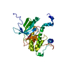


 PDBj
PDBj Assembly
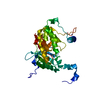


 Mass: 65.409 Da / Num. of mol.: 1 / Source method: obtained synthetically / Formula: Zn
 Mass: 18.015 Da / Num. of mol.: 18 / Source method: isolated from a natural source / Formula: H2O
 Sample preparation
 / Beamline: BW6 / Wavelength: 1.05 Å
 Processing