 Movie
Movie Controller

[English] 日本語
 Yorodumi
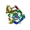
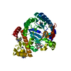
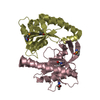
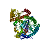
- PDB-2a6p: Structure Solution to 2.2 Angstrom and Functional Characterisatio... -
+ Open data

- Basic information
| Entry | Database: PDB / ID: 2a6p | ||||||
|---|---|---|---|---|---|---|---|
| Title | Structure Solution to 2.2 Angstrom and Functional Characterisation of the Open Reading Frame Rv3214 from Mycobacterium tuberculosis | ||||||
 Components Components | POSSIBLE PHOSPHOGLYCERATE MUTASE GPM2 | ||||||
 Keywords Keywords | STRUCTURAL GENOMICS / UNKNOWN FUNCTION / predicted phosphoglycerate mutase / PSI / Protein Structure Initiative / TB Structural Genomics Consortium / TBSGC | ||||||
| Function / homology |  Function and homology information Function and homology informationprotein histidine phosphatase activity / regulation of phosphorelay signal transduction system / acid phosphatase / acid phosphatase activity / fructose-bisphosphatase / fructose 1,6-bisphosphate 1-phosphatase activity / gluconeogenesis / protein homodimerization activity Similarity search - Function | ||||||
| Biological species |   Mycobacterium tuberculosis (bacteria) Mycobacterium tuberculosis (bacteria) | ||||||
| Method |  X-RAY DIFFRACTION / SYNCHROTRON / MAD / Resolution: 2.2 Å X-RAY DIFFRACTION / SYNCHROTRON / MAD / Resolution: 2.2 Å | ||||||
 Authors Authors | Watkins, H.A. / Yu, M. / Baker, E.N. / TB Structural Genomics Consortium (TBSGC) | ||||||
 Citation Citation | Journal: J.Bacteriol. / Year: 2006 Title: Structural and Functional Analysis of Rv3214 from Mycobacterium tuberculosis, a Protein with Conflicting Functional Annotations, Leads to Its Characterization as a Phosphatase. Authors: Watkins, H.A. / Baker, E.N. | ||||||
| History |
|
- Structure visualization
| Structure viewer | Molecule: MolmilJmol/JSmol |
|---|
- Downloads & links
-Download
| PDBx/mmCIF format | 2a6p.cif.gz | 87.1 KB | Display | PDBx/mmCIF format |
|---|---|---|---|---|
| PDB format | pdb2a6p.ent.gz | 66.1 KB | Display | PDB format |
| PDBx/mmJSON format | 2a6p.json.gz | Tree view | PDBx/mmJSON format | |
| Others |  Other downloads Other downloads |
-Validation report
| Summary document | 2a6p_validation.pdf.gz | 468.4 KB | Display | wwPDB validaton report |
|---|---|---|---|---|
| Full document | 2a6p_full_validation.pdf.gz | 481.8 KB | Display | |
| Data in XML | 2a6p_validation.xml.gz | 18.7 KB | Display | |
| Data in CIF | 2a6p_validation.cif.gz | 24.9 KB | Display | |
| Arichive directory | https://data.pdbj.org/pub/pdb/validation_reports/a6/2a6pftp://data.pdbj.org/pub/pdb/validation_reports/a6/2a6p | HTTPS FTP |
-Related structure data
| Similar structure data | |
|---|---|
| Other databases |


-Links
 PDBj
PDBj


- Assembly
| Deposited unit | 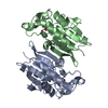
| ||||||||
|---|---|---|---|---|---|---|---|---|---|
| 1 |
| ||||||||
| Unit cell |
| ||||||||
| Details | Biological Unit is the dimer found in the assymetric unit |
-Components
| #1: Protein | Mass: 22432.316 Da / Num. of mol.: 2 Source method: isolated from a genetically manipulated source Source: (gene. exp.) Mycobacterium tuberculosis (bacteria) / Strain: H37Rv / Gene: Rv3214 (EntD) / Plasmid: pPROEX HTa / Species (production host): Escherichia coli / Production host: References: UniProt: Q6MWZ7, UniProt: Q7D5X2*PLUS, EC: 5.4.2.1 #2: Chemical |   Mass: 96.063 Da / Num. of mol.: 2 / Source method: obtained synthetically / Formula: SO4 Mass: 96.063 Da / Num. of mol.: 2 / Source method: obtained synthetically / Formula: SO4#3: Chemical |   Mass: 92.094 Da / Num. of mol.: 2 / Source method: obtained synthetically / Formula: C3H8O3 Mass: 92.094 Da / Num. of mol.: 2 / Source method: obtained synthetically / Formula: C3H8O3#4: Water | ChemComp-HOH / |  Mass: 18.015 Da / Num. of mol.: 106 / Source method: isolated from a natural source / Formula: H2O Mass: 18.015 Da / Num. of mol.: 106 / Source method: isolated from a natural source / Formula: H2O |
|---|
-Experimental details
-Experiment
| Experiment | Method: X-RAY DIFFRACTION / Number of used crystals: 1 |
|---|
- Sample preparation
| Crystal | Density Matthews: 2 Å3/Da / Density % sol: 37.7 % |
|---|---|
| Crystal grow | Temperature: 291 K / Method: vapor diffusion, sitting drop / pH: 7 Details: MPD, Lithium sulphate, Imidazole-HCl, pH 7.0, VAPOR DIFFUSION, SITTING DROP, temperature 291K |
-Data collection
| Diffraction | Mean temperature: 113 K | |||||||||
|---|---|---|---|---|---|---|---|---|---|---|
| Diffraction source | Source: SYNCHROTRON / Site: ALS  / Beamline: 5.0.2 / Wavelength: 0.91840, 0.97972 / Beamline: 5.0.2 / Wavelength: 0.91840, 0.97972 | |||||||||
| Radiation | Protocol: MAD / Monochromatic (M) / Laue (L): M / Scattering type: x-ray | |||||||||
| Radiation wavelength |
| |||||||||
| Reflection | Resolution: 2.15→50 Å / Num. obs: 22262 / Biso Wilson estimate: 10.8 Å2 / Rmerge(I) obs: 0.105 / Net I/σ(I): 57.3 | |||||||||
| Reflection shell | Resolution: 2.15→2.25 Å / Rmerge(I) obs: 0.157 / Mean I/σ(I) obs: 10.1 / Num. unique all: 10306 / % possible all: 96.2 |
- Processing
| Software |
| ||||||||||||||||||||||||||||||||||||
|---|---|---|---|---|---|---|---|---|---|---|---|---|---|---|---|---|---|---|---|---|---|---|---|---|---|---|---|---|---|---|---|---|---|---|---|---|---|
| Refinement | Method to determine structure: MAD / Resolution: 2.2→28.9 Å / Rfactor Rfree error: 0.008 / Data cutoff high absF: 1373222.55 / Data cutoff low absF: 0 / Isotropic thermal model: RESTRAINED / Cross valid method: THROUGHOUT / σ(F): 0
| ||||||||||||||||||||||||||||||||||||
| Solvent computation | Solvent model: FLAT MODEL / Bsol: 39.5739 Å2 / ksol: 0.352897 e/Å3 | ||||||||||||||||||||||||||||||||||||
| Displacement parameters | Biso mean: 25.4 Å2
| ||||||||||||||||||||||||||||||||||||
| Refine analyze |
| ||||||||||||||||||||||||||||||||||||
| Refinement step | Cycle: LAST / Resolution: 2.2→28.9 Å
| ||||||||||||||||||||||||||||||||||||
| Refine LS restraints |
| ||||||||||||||||||||||||||||||||||||
| Refine LS restraints NCS | NCS model details: CONSTR | ||||||||||||||||||||||||||||||||||||
| LS refinement shell | Resolution: 2.2→2.34 Å / Rfactor Rfree error: 0.023 / Total num. of bins used: 6
| ||||||||||||||||||||||||||||||||||||
| Xplor file |
|
