 Movie
Movie Controller

[English] 日本語
 Yorodumi
- PDB-2a2g: THE CRYSTAL STRUCTURES OF A2U-GLOBULIN AND ITS COMPLEX WITH A HYA... -
+ Open data

- Basic information
| Entry | Database: PDB / ID: 2a2g | ||||||
|---|---|---|---|---|---|---|---|
| Title | THE CRYSTAL STRUCTURES OF A2U-GLOBULIN AND ITS COMPLEX WITH A HYALINE DROPLET INDUCER. | ||||||
 Components Components | PROTEIN (ALPHA-2U-GLOBULIN) | ||||||
 Keywords Keywords | LIPID BINDING PROTEIN / LIPID-BINDING PROTEIN | ||||||
| Function / homology |  Function and homology information Function and homology informationpheromone binding / negative regulation of lipid biosynthetic process / positive regulation of glucose metabolic process / odorant binding / energy reserve metabolic process / insulin receptor activity / negative regulation of insulin secretion involved in cellular response to glucose stimulus / heat generation / cellular response to lipid / positive regulation of lipid metabolic process ...pheromone binding / negative regulation of lipid biosynthetic process / positive regulation of glucose metabolic process / odorant binding / energy reserve metabolic process / insulin receptor activity / negative regulation of insulin secretion involved in cellular response to glucose stimulus / heat generation / cellular response to lipid / positive regulation of lipid metabolic process / locomotor rhythm / small molecule binding / negative regulation of lipid storage / negative regulation of gluconeogenesis / aerobic respiration / mitochondrion organization / glucose homeostasis / positive regulation of phosphatidylinositol 3-kinase/protein kinase B signal transduction / negative regulation of DNA-templated transcription / positive regulation of gene expression / : / nucleus / cytosol Similarity search - Function | ||||||
| Biological species |  | ||||||
| Method |  X-RAY DIFFRACTION / MOLECULAR REPLACEMENT / Resolution: 2.9 Å X-RAY DIFFRACTION / MOLECULAR REPLACEMENT / Resolution: 2.9 Å | ||||||
 Authors Authors | Chaudhuri, B.N. / Kleywegt, G.J. / Jones, T.A. | ||||||
 Citation Citation | Journal: Acta Crystallogr.,Sect.D / Year: 1999 Title: The structures of alpha 2u-globulin and its complex with a hyaline droplet inducer. Authors: Chaudhuri, B.N. / Kleywegt, G.J. / Bjorkman, J. / Lehman-McKeeman, L.D. / Oliver, J.D. / Jones, T.A. #1: Journal: Adv.Protein Chem. / Year: 1994Title: Lipid-Binding Proteins: A Family of Fatty Acid and Retinoid Transport Proteins Authors: Banaszak, L. / Winter, N. / Xu, Z. / Bernlohr, D.A. / Cowan, S.W. / Jones, T.A. #2: Journal: Nature / Year: 1992Title: Pheromone Binding to Two Rodent Urinary Proteins Revealed by X-Ray Crystallography Authors: Bocskei, Z. / Groom, C.R. / Flower, D.R. / Wright, C.E. / Phillips, S.E.V. / Cavaggioni, A. / Findlay, J.B.C. / North, A.C.T. | ||||||
| History |
|
- Structure visualization
| Structure viewer | Molecule: MolmilJmol/JSmol |
|---|
- Downloads & links
-Download
| PDBx/mmCIF format | 2a2g.cif.gz | 127.4 KB | Display | PDBx/mmCIF format |
|---|---|---|---|---|
| PDB format | pdb2a2g.ent.gz | 102.2 KB | Display | PDB format |
| PDBx/mmJSON format | 2a2g.json.gz | Tree view | PDBx/mmJSON format | |
| Others |  Other downloads Other downloads |
-Validation report
| Arichive directory | https://data.pdbj.org/pub/pdb/validation_reports/a2/2a2gftp://data.pdbj.org/pub/pdb/validation_reports/a2/2a2g | HTTPS FTP |
|---|
-Related structure data


-Links
 PDBj
PDBj

- Assembly
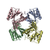
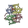
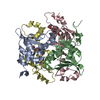
| Deposited unit | 
| ||||||||||||||||
|---|---|---|---|---|---|---|---|---|---|---|---|---|---|---|---|---|---|
| 1 |
| ||||||||||||||||
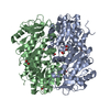
| Unit cell |
| ||||||||||||||||
| Noncrystallographic symmetry (NCS) | NCS oper:
|
-Components
| #1: Protein | Mass: 20764.854 Da / Num. of mol.: 4 / Source method: isolated from a natural source / Details: PURIFIED FROM MALE RAT URINE / Source: (natural) #2: Chemical | ChemComp-LEO / 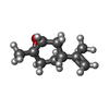  Mass: 152.233 Da / Num. of mol.: 4 / Source method: obtained synthetically / Formula: C10H16O Mass: 152.233 Da / Num. of mol.: 4 / Source method: obtained synthetically / Formula: C10H16OHas protein modification | Y | |
|---|
-Experimental details
-Experiment
| Experiment | Method: X-RAY DIFFRACTION / Number of used crystals: 2 |
|---|
- Sample preparation
| Crystal | Density Matthews: 2.5 Å3/Da / Density % sol: 51 % | |||||||||||||||||||||||||
|---|---|---|---|---|---|---|---|---|---|---|---|---|---|---|---|---|---|---|---|---|---|---|---|---|---|---|
| Crystal grow | pH: 4.8 / Details: pH 4.80 | |||||||||||||||||||||||||
| Crystal | *PLUS | |||||||||||||||||||||||||
| Crystal grow | *PLUS Temperature: 277 K / Method: vapor diffusion, sitting drop | |||||||||||||||||||||||||
| Components of the solutions | *PLUS
|
-Data collection
| Diffraction | Mean temperature: 277 K |
|---|---|
| Diffraction source | Source: ROTATING ANODE / Type: RIGAKU / Wavelength: 1.5418 |
| Detector | Type: RIGAKU RAXIS IIC / Detector: IMAGE PLATE |
| Radiation | Monochromator: GRAPHITE MONOCHROMATOR / Protocol: SINGLE WAVELENGTH / Monochromatic (M) / Laue (L): M / Scattering type: x-ray |
| Radiation wavelength | Wavelength: 1.5418 Å / Relative weight: 1 |
| Reflection | Resolution: 2.9→49 Å / Num. obs: 15350 / % possible obs: 88 % / Redundancy: 3.2 % / Rmerge(I) obs: 0.102 / Net I/σ(I): 11.3 |
| Reflection shell | Resolution: 2.9→2.98 Å / Redundancy: 3.1 % / Rmerge(I) obs: 0.35 / Mean I/σ(I) obs: 3.6 / % possible all: 91.3 |
| Reflection | *PLUS Highest resolution: 2.9 Å / Lowest resolution: 49 Å / Redundancy: 3.2 % / Num. measured all: 49636 |
| Reflection shell | *PLUS % possible obs: 91.3 % / Redundancy: 3.1 % / Mean I/σ(I) obs: 3.6 |
- Processing
| Software |
| ||||||||||||||||||||||||||||||||||||||||||||||||||||||||||||
|---|---|---|---|---|---|---|---|---|---|---|---|---|---|---|---|---|---|---|---|---|---|---|---|---|---|---|---|---|---|---|---|---|---|---|---|---|---|---|---|---|---|---|---|---|---|---|---|---|---|---|---|---|---|---|---|---|---|---|---|---|---|
| Refinement | Method to determine structure: MOLECULAR REPLACEMENT Starting model: AN ORTHORHOMBIC FORM OF A2U Resolution: 2.9→10 Å / Rfactor Rfree error: 0.007 / Cross valid method: THROUGHOUT / σ(F): 0 / Details: THE LIGAND WAS *NOT* INCLUDED IN THE REFINEMENT
| ||||||||||||||||||||||||||||||||||||||||||||||||||||||||||||
| Solvent computation | Solvent model: FLAT MODEL | ||||||||||||||||||||||||||||||||||||||||||||||||||||||||||||
| Displacement parameters | Biso mean: 41.4 Å2 | ||||||||||||||||||||||||||||||||||||||||||||||||||||||||||||
| Refine analyze | Luzzati coordinate error obs: 0.4 Å / Luzzati d res low obs: 5 Å / Luzzati sigma a obs: 0.45 Å | ||||||||||||||||||||||||||||||||||||||||||||||||||||||||||||
| Refinement step | Cycle: LAST / Resolution: 2.9→10 Å
| ||||||||||||||||||||||||||||||||||||||||||||||||||||||||||||
| Refine LS restraints |
| ||||||||||||||||||||||||||||||||||||||||||||||||||||||||||||
| Refine LS restraints NCS | NCS model details: CONSTRAINED | ||||||||||||||||||||||||||||||||||||||||||||||||||||||||||||
| LS refinement shell | Resolution: 2.9→3.08 Å / Rfactor Rfree error: 0.03 / Total num. of bins used: 6
| ||||||||||||||||||||||||||||||||||||||||||||||||||||||||||||
| Xplor file | Serial no: 1 / Param file: PROTEIN_REP.PARAM / Topol file: PROTEIN.TOP | ||||||||||||||||||||||||||||||||||||||||||||||||||||||||||||
| Software | *PLUS Name: CNS / Version: 0.3 / Classification: refinement | ||||||||||||||||||||||||||||||||||||||||||||||||||||||||||||
| Refinement | *PLUS Rfactor Rfree: 0.26 | ||||||||||||||||||||||||||||||||||||||||||||||||||||||||||||
| Solvent computation | *PLUS | ||||||||||||||||||||||||||||||||||||||||||||||||||||||||||||
| Displacement parameters | *PLUS | ||||||||||||||||||||||||||||||||||||||||||||||||||||||||||||
| Refine LS restraints | *PLUS
| ||||||||||||||||||||||||||||||||||||||||||||||||||||||||||||
| LS refinement shell | *PLUS Rfactor Rfree: 0.398 / Rfactor Rwork: 0.321 |
