Method to determine structure: SAD / Resolution: 1.801→64.19 Å / Cor.coef. Fo:Fc: 0.957 / Cor.coef. Fo:Fc free: 0.949 / SU R Cruickshank DPI: 0.108 / Cross valid method: THROUGHOUT / SU R Blow DPI: 0.119 / SU Rfree Blow DPI: 0.107 / SU Rfree Cruickshank DPI: 0.101
Rfactor
Num. reflection
% reflection
Selection details
Rfree
0.1871
1922
-
RANDOM
Rwork
0.1606
26597
-
-
obs
0.1624
28519
99.1 %
-
Displacement parameters
Biso mean: 20.3 Å2
Baniso -1
Baniso -2
Baniso -3
1-
-2.1618 Å2
0 Å2
-3.1949 Å2
2-
-
1.2767 Å2
0 Å2
3-
-
-
0.8851 Å2
Refine analyze
Luzzati coordinate error obs: 0.17 Å
Refinement step
Cycle: LAST / Resolution: 1.801→64.19 Å
Protein
Nucleic acid
Ligand
Solvent
Total
Num. atoms
2176
0
14
362
2552
Refine LS restraints
Refine-ID
Type
Dev ideal
Number
Restraint function
Weight
X-RAY DIFFRACTION
t_bond_d
0.009
2255
HARMONIC
2
X-RAY DIFFRACTION
t_angle_deg
0.91
3066
HARMONIC
2
X-RAY DIFFRACTION
t_dihedral_angle_d
748
SINUSOIDAL
2
X-RAY DIFFRACTION
t_gen_planes
391
HARMONIC
5
X-RAY DIFFRACTION
t_it
2255
HARMONIC
10
X-RAY DIFFRACTION
t_chiral_improper_torsion
278
SEMIHARMONIC
5
X-RAY DIFFRACTION
t_ideal_dist_contact
2245
SEMIHARMONIC
4
X-RAY DIFFRACTION
t_omega_torsion
4.02
X-RAY DIFFRACTION
t_other_torsion
15.09
LS refinement shell
Resolution: 1.801→1.82 Å
Rfactor
Num. reflection
% reflection
Rfree
0.1857
37
-
Rwork
0.1735
534
-
obs
-
-
84.68 %
Refinement TLS params.
Refine-ID: X-RAY DIFFRACTION
ID
L11 (°2)
L12 (°2)
L13 (°2)
L22 (°2)
L23 (°2)
L33 (°2)
S11 (Å °)
S12 (Å °)
S13 (Å °)
S21 (Å °)
S22 (Å °)
S23 (Å °)
S31 (Å °)
S32 (Å °)
S33 (Å °)
T11 (Å2)
T12 (Å2)
T13 (Å2)
T22 (Å2)
T23 (Å2)
T33 (Å2)
Origin x (Å)
Origin y (Å)
Origin z (Å)
1
0.3678
-0.1114
-0.0508
0.7077
-0.0069
0.1424
0.002
0.0169
0.0917
0.0153
0.0463
0.0287
0.0518
0.027
-0.0484
-0.0254
0.006
0.0019
-0.0141
0.0222
0.0117
22.784
1.422
49.7252
2
0.7209
0.4782
-0.2678
0.5254
-0.1003
0.4147
-0.0218
0.0277
0.0683
0.0096
0.0329
-0.0083
0.0288
-0.0483
-0.0111
-0.0212
0.0065
0.0017
-0.0177
-0.0006
0.0022
12.1001
29.4262
46.2169
Refinement TLS group
ID
Refine-ID
Refine TLS-ID
Selection details
Auth asym-ID
Auth seq-ID
1
X-RAY DIFFRACTION
1
{ A|* }
A
1 - 141
2
X-RAY DIFFRACTION
1
{ A|* }
A
201
3
X-RAY DIFFRACTION
2
{ B|* }
B
1 - 141
4
X-RAY DIFFRACTION
2
{ B|* }
B
201
+
About Yorodumi
-
News
-
Feb 9, 2022. New format data for meta-information of EMDB entries
New format data for meta-information of EMDB entries
Version 3 of the EMDB header file is now the official format.
The previous official version 1.9 will be removed from the archive.
In the structure databanks used in Yorodumi, some data are registered as the other names, "COVID-19 virus" and "2019-nCoV". Here are the details of the virus and the list of structure data.
Jan 31, 2019. EMDB accession codes are about to change! (news from PDBe EMDB page)
EMDB accession codes are about to change! (news from PDBe EMDB page)
The allocation of 4 digits for EMDB accession codes will soon come to an end. Whilst these codes will remain in use, new EMDB accession codes will include an additional digit and will expand incrementally as the available range of codes is exhausted. The current 4-digit format prefixed with “EMD-” (i.e. EMD-XXXX) will advance to a 5-digit format (i.e. EMD-XXXXX), and so on. It is currently estimated that the 4-digit codes will be depleted around Spring 2019, at which point the 5-digit format will come into force.
The EM Navigator/Yorodumi systems omit the EMD- prefix.
Related info.:Q: What is EMD? / ID/Accession-code notation in Yorodumi/EM Navigator
Yorodumi is a browser for structure data from EMDB, PDB, SASBDB, etc.
This page is also the successor to EM Navigator detail page, and also detail information page/front-end page for Omokage search.
The word "yorodu" (or yorozu) is an old Japanese word meaning "ten thousand". "mi" (miru) is to see.
Related info.:EMDB / PDB / SASBDB / Comparison of 3 databanks / Yorodumi Search / Aug 31, 2016. New EM Navigator & Yorodumi / Yorodumi Papers / Jmol/JSmol / Function and homology information / Changes in new EM Navigator and Yorodumi
 Movie
Movie Controller

 Yorodumi
 Open data
 Basic information
 Components
 Keywords
 Function and homology information
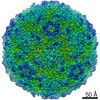 Coxsackievirus A16
 X-RAY DIFFRACTION /
  Authors
 United Kingdom, 1items
  Citation
 Structure visualization
 Downloads & links
 Other downloads
 PDBj
PDBj

 Assembly


 Mass: 92.094 Da / Num. of mol.: 2 / Source method: obtained synthetically / Formula: C3H8O3
 Mass: 65.409 Da / Num. of mol.: 2 / Source method: obtained synthetically / Formula: Zn
 Mass: 18.015 Da / Num. of mol.: 362 / Source method: isolated from a natural source / Formula: H2O
 Sample preparation
 Processing