 Movie
Movie Controller

[English] 日本語
 Yorodumi
- PDB-28ub: LASV cap-snatching endonuclease in complex with 2,4-dioxo-4-pheny... -
+ Open data

- Basic information
| Entry | Database: PDB / ID: 28ub | |||||||||||||||||||||
|---|---|---|---|---|---|---|---|---|---|---|---|---|---|---|---|---|---|---|---|---|---|---|
| Title | LASV cap-snatching endonuclease in complex with 2,4-dioxo-4-phenylbutanoic acid - serial synchrotron crystallography | |||||||||||||||||||||
 Components Components | RNA-directed RNA polymerase L | |||||||||||||||||||||
 Keywords Keywords | HYDROLASE / drug development / Lassa hemorrhagic fever / L protein / Arenaviridae | |||||||||||||||||||||
| Function / homology |  Function and homology information Function and homology informationnegative stranded viral RNA replication / cap snatching / hydrolase activity / host cell / Hydrolases; Acting on ester bonds / nucleotide binding / RNA-directed RNA polymerase / RNA-directed RNA polymerase activity / host cell cytoplasm / metal ion binding Similarity search - Function | |||||||||||||||||||||
| Biological species |  Mammarenavirus lassaense Mammarenavirus lassaense | |||||||||||||||||||||
| Method |  X-RAY DIFFRACTION / SYNCHROTRON / MOLECULAR REPLACEMENT / Resolution: 1.66 Å X-RAY DIFFRACTION / SYNCHROTRON / MOLECULAR REPLACEMENT / Resolution: 1.66 Å | |||||||||||||||||||||
 Authors Authors | Falke, S. / Reinke, P.Y.A. / Senst, J.M. / Lewe, P. / Witt, S. / Meents, A. / Guenther, S. | |||||||||||||||||||||
| Funding support |  Germany, 6items Germany, 6items
| |||||||||||||||||||||
 Citation Citation | Journal: Acta Crystallogr D Struct Biol / Year: 2026 Title: Perspectives for pharmaceutical screening at XFEL sources using the example of Lassa virus endonuclease. Authors: Falke, S. / Reinke, P.Y.A. / Rosenberg, D. / Fischer, P. / Meyer, J. / Galchenkova, M. / Tolstikova, A. / Mariani, V. / Senst, J.M. / Lewe, P. / Witt, S. / Wagner, A. / Chapman, H.N. / ...Authors: Falke, S. / Reinke, P.Y.A. / Rosenberg, D. / Fischer, P. / Meyer, J. / Galchenkova, M. / Tolstikova, A. / Mariani, V. / Senst, J.M. / Lewe, P. / Witt, S. / Wagner, A. / Chapman, H.N. / Hunter, M. / Gunther, S. / Meents, A. | |||||||||||||||||||||
| History |
|
- Structure visualization
| Structure viewer | Molecule: MolmilJmol/JSmol |
|---|
- Downloads & links
-Download
| PDBx/mmCIF format | 28ub.cif.gz | 55.4 KB | Display | PDBx/mmCIF format |
|---|---|---|---|---|
| PDB format | pdb28ub.ent.gz | 36.5 KB | Display | PDB format |
| PDBx/mmJSON format | 28ub.json.gz | Tree view | PDBx/mmJSON format | |
| Others |  Other downloads Other downloads |
-Validation report
| Arichive directory | https://data.pdbj.org/pub/pdb/validation_reports/8u/28ubftp://data.pdbj.org/pub/pdb/validation_reports/8u/28ub | HTTPS FTP |
|---|
-Related structure data
| Related structure data |  28jgC  28jhC  28jiC  28uaC  28ucC  29ikC  29ilC  29imC C: citing same article ( |
|---|---|
| Similar structure data |
-Links
 PDBj
PDBj- Assembly
| Deposited unit | 
| ||||||||||||
|---|---|---|---|---|---|---|---|---|---|---|---|---|---|
| 1 |
| ||||||||||||
| Unit cell |
|
-Components
| #1: Protein | Mass: 22922.141 Da / Num. of mol.: 1 Source method: isolated from a genetically manipulated source Source: (gene. exp.) Mammarenavirus lassaense / Production host:   Escherichia coli BL21(DE3) (bacteria) Escherichia coli BL21(DE3) (bacteria)References: UniProt: Q6GWS2, RNA-directed RNA polymerase, Hydrolases; Acting on ester bonds | ||||||||||
|---|---|---|---|---|---|---|---|---|---|---|---|
| #2: Chemical |   Mass: 24.305 Da / Num. of mol.: 2 / Source method: obtained synthetically / Formula: Mg Mass: 24.305 Da / Num. of mol.: 2 / Source method: obtained synthetically / Formula: Mg#3: Chemical | ChemComp-XI7 / | 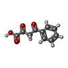  Mass: 192.168 Da / Num. of mol.: 1 / Source method: obtained synthetically / Formula: C10H8O4 / Feature type: SUBJECT OF INVESTIGATION Mass: 192.168 Da / Num. of mol.: 1 / Source method: obtained synthetically / Formula: C10H8O4 / Feature type: SUBJECT OF INVESTIGATION#4: Chemical | ChemComp-DMS / | 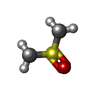 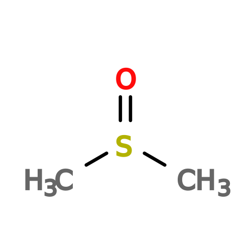 Mass: 78.133 Da / Num. of mol.: 1 / Source method: obtained synthetically / Formula: C2H6OS / Comment: DMSO, precipitant*YM Mass: 78.133 Da / Num. of mol.: 1 / Source method: obtained synthetically / Formula: C2H6OS / Comment: DMSO, precipitant*YM#5: Water | ChemComp-HOH / |  Mass: 18.015 Da / Num. of mol.: 80 / Source method: isolated from a natural source / Formula: H2O Mass: 18.015 Da / Num. of mol.: 80 / Source method: isolated from a natural source / Formula: H2OHas ligand of interest | Y | Has protein modification | Y | |
-Experimental details
-Experiment
| Experiment | Method: X-RAY DIFFRACTION / Number of used crystals: 1 |
|---|
- Sample preparation
| Crystal | Density Matthews: 2.51 Å3/Da / Density % sol: 50.97 % |
|---|---|
| Crystal grow | Temperature: 293 K / Method: batch mode Details: After limited proteolysis with subtilisin A, 7.5 mg/ml target protein were mixed with an equal volume of crystallization solution (10% w/v PEG 10000, 250 mM MgSO4 and 100 mM Tris/HCl pH 9.0) |
-Data collection
| Diffraction | Mean temperature: 293 K / Serial crystal experiment: N |
|---|---|
| Diffraction source | Source: SYNCHROTRON / Site: PETRA III, DESY / Beamline: P09 HiPhaX / Wavelength: 0.7749 Å |
| Detector | Type: DECTRIS PILATUS 6M / Detector: PIXEL / Date: Dec 22, 2024 |
| Radiation | Protocol: SINGLE WAVELENGTH / Monochromatic (M) / Laue (L): M / Scattering type: x-ray |
| Radiation wavelength | Wavelength: 0.7749 Å / Relative weight: 1 |
| Reflection | Resolution: 1.66→45.01 Å / Num. obs: 29030 / % possible obs: 100 % / Redundancy: 197.8 % / Biso Wilson estimate: 22.55 Å2 / CC1/2: 0.994 / CC star: 0.998 / Net I/σ(I): 8.3 |
| Reflection shell | Resolution: 1.66→1.69 Å / Mean I/σ(I) obs: 1 / Num. unique obs: 1404 / CC1/2: 0.52 / CC star: 0.827 |
- Processing
| Software |
| |||||||||||||||||||||||||||||||||||||||||||||||||||||||||||||||||||||||||||||||||||||||||||||||||||||||||
|---|---|---|---|---|---|---|---|---|---|---|---|---|---|---|---|---|---|---|---|---|---|---|---|---|---|---|---|---|---|---|---|---|---|---|---|---|---|---|---|---|---|---|---|---|---|---|---|---|---|---|---|---|---|---|---|---|---|---|---|---|---|---|---|---|---|---|---|---|---|---|---|---|---|---|---|---|---|---|---|---|---|---|---|---|---|---|---|---|---|---|---|---|---|---|---|---|---|---|---|---|---|---|---|---|---|---|
| Refinement | Method to determine structure: MOLECULAR REPLACEMENT / Resolution: 1.66→44.16 Å / SU ML: 0.2348 / Cross valid method: FREE R-VALUE / σ(F): 1.33 / Phase error: 22.325 Stereochemistry target values: GeoStd + Monomer Library + CDL v1.2
| |||||||||||||||||||||||||||||||||||||||||||||||||||||||||||||||||||||||||||||||||||||||||||||||||||||||||
| Solvent computation | Shrinkage radii: 0.9 Å / VDW probe radii: 1.1 Å / Solvent model: FLAT BULK SOLVENT MODEL | |||||||||||||||||||||||||||||||||||||||||||||||||||||||||||||||||||||||||||||||||||||||||||||||||||||||||
| Displacement parameters | Biso mean: 29.95 Å2 | |||||||||||||||||||||||||||||||||||||||||||||||||||||||||||||||||||||||||||||||||||||||||||||||||||||||||
| Refinement step | Cycle: LAST / Resolution: 1.66→44.16 Å
| |||||||||||||||||||||||||||||||||||||||||||||||||||||||||||||||||||||||||||||||||||||||||||||||||||||||||
| Refine LS restraints |
| |||||||||||||||||||||||||||||||||||||||||||||||||||||||||||||||||||||||||||||||||||||||||||||||||||||||||
| LS refinement shell |
|
