 Movie
Movie Controller

[English] 日本語
 Yorodumi
- PDB-28mg: crystal structure of Dpo31, of a tail-spike protein with depolyme... -
+ Open data

- Basic information
| Entry | Database: PDB / ID: 28mg | ||||||
|---|---|---|---|---|---|---|---|
| Title | crystal structure of Dpo31, of a tail-spike protein with depolymerase activity identified in a marine podovirus | ||||||
 Components Components | Dpo31-deltaD1, viral depolymerase depleted of domain 1 | ||||||
 Keywords Keywords | VIRAL PROTEIN / tailspike protein / marine virus / podovirus | ||||||
| Function / homology | DI(HYDROXYETHYL)ETHER Function and homology information Function and homology information | ||||||
| Biological species |  Cobetia phage Carin1 (virus) Cobetia phage Carin1 (virus) | ||||||
| Method |  X-RAY DIFFRACTION / SYNCHROTRON / MOLECULAR REPLACEMENT / Resolution: 2.15 Å X-RAY DIFFRACTION / SYNCHROTRON / MOLECULAR REPLACEMENT / Resolution: 2.15 Å | ||||||
 Authors Authors | Czjzek, M. / Sirigu, S. / Roret, T. / Baudoux, A.C. | ||||||
| Funding support |  France, 1items France, 1items
| ||||||
 Citation Citation | Journal: Acta Crystallogr D Struct Biol / Year: 2026 Title: Biochemical and structural characterization of a tail-spike protein with depolymerase activity identified in a marine podovirus. Authors: Sirigu, S. / Roret, T. / Mocaer, P.Y. / Larocque, R. / Jouanneau, D. / Legrand, P. / Baudoux, A.C. / Czjzek, M. | ||||||
| History |
|
- Structure visualization
| Structure viewer | Molecule: MolmilJmol/JSmol |
|---|
- Downloads & links
-Download
| PDBx/mmCIF format | 28mg.cif.gz | 640.4 KB | Display | PDBx/mmCIF format |
|---|---|---|---|---|
| PDB format | pdb28mg.ent.gz | 523.7 KB | Display | PDB format |
| PDBx/mmJSON format | 28mg.json.gz | Tree view | PDBx/mmJSON format | |
| Others |  Other downloads Other downloads |
-Validation report
| Arichive directory | https://data.pdbj.org/pub/pdb/validation_reports/8m/28mgftp://data.pdbj.org/pub/pdb/validation_reports/8m/28mg | HTTPS FTP |
|---|
-Related structure data


-Links
 PDBj
PDBj- Assembly
| Deposited unit | 
| ||||||||
|---|---|---|---|---|---|---|---|---|---|
| 1 |
| ||||||||
| Unit cell |
|
-Components
| #1: Protein | Mass: 86858.156 Da / Num. of mol.: 3 Source method: isolated from a genetically manipulated source Details: The crystal structure contains a truncated version missing domain 1 of the polypetidic chain : Dpo31-DeltaD1. The crystal structure also covers the mature protein only, and does not contain ...Details: The crystal structure contains a truncated version missing domain 1 of the polypetidic chain : Dpo31-DeltaD1. The crystal structure also covers the mature protein only, and does not contain the C-terminal domain D4. Source: (gene. exp.) Cobetia phage Carin1 (virus) / Production host:   Escherichia coli BL21(DE3) (bacteria) Escherichia coli BL21(DE3) (bacteria)#2: Chemical | ChemComp-GOL / 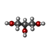 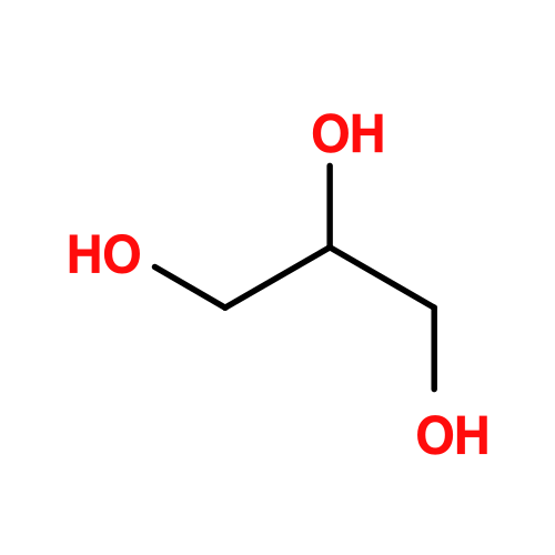 Mass: 92.094 Da / Num. of mol.: 4 / Source method: obtained synthetically / Formula: C3H8O3 Mass: 92.094 Da / Num. of mol.: 4 / Source method: obtained synthetically / Formula: C3H8O3#3: Chemical |   Mass: 24.305 Da / Num. of mol.: 3 / Source method: obtained synthetically / Formula: Mg Mass: 24.305 Da / Num. of mol.: 3 / Source method: obtained synthetically / Formula: Mg#4: Chemical | ChemComp-PEG / | 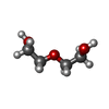 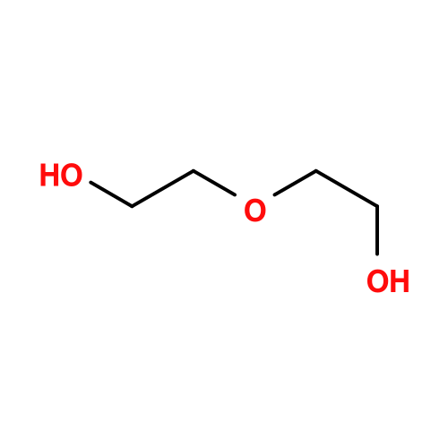 Mass: 106.120 Da / Num. of mol.: 1 / Source method: obtained synthetically / Formula: C4H10O3 Mass: 106.120 Da / Num. of mol.: 1 / Source method: obtained synthetically / Formula: C4H10O3#5: Water | ChemComp-HOH / |  Mass: 18.015 Da / Num. of mol.: 411 / Source method: isolated from a natural source / Formula: H2O Mass: 18.015 Da / Num. of mol.: 411 / Source method: isolated from a natural source / Formula: H2OHas ligand of interest | N | Has protein modification | N | |
|---|
-Experimental details
-Experiment
| Experiment | Method: X-RAY DIFFRACTION / Number of used crystals: 1 |
|---|
- Sample preparation
| Crystal | Density Matthews: 2.51 Å3/Da / Density % sol: 50.99 % |
|---|---|
| Crystal grow | Temperature: 283 K / Method: vapor diffusion, hanging drop / pH: 7.4 Details: 20% PEG 3350 and 200 mM Potassium nitrate. drop ratio 2:1 (protein to reservoir) |
-Data collection
| Diffraction | Mean temperature: 100 K / Serial crystal experiment: N |
|---|---|
| Diffraction source | Source: SYNCHROTRON / Site: SOLEIL / Beamline: PROXIMA 2 / Wavelength: 0.979 Å |
| Detector | Type: DECTRIS PILATUS 6M / Detector: PIXEL / Date: Feb 5, 2022 |
| Radiation | Protocol: SINGLE WAVELENGTH / Monochromatic (M) / Laue (L): M / Scattering type: x-ray |
| Radiation wavelength | Wavelength: 0.979 Å / Relative weight: 1 |
| Reflection | Resolution: 2.15→47.4 Å / Num. obs: 59091 / % possible obs: 63.4 % / Redundancy: 6.2 % / Biso Wilson estimate: 26.77 Å2 / CC1/2: 0.997 / Rmerge(I) obs: 0.115 / Rpim(I) all: 0.05 / Net I/σ(I): 9.5 |
| Reflection shell | Resolution: 2.15→2.21 Å / Redundancy: 2.3 % / Rmerge(I) obs: 0.543 / Num. unique obs: 1200 / CC1/2: 0.613 / Rpim(I) all: 0.351 / % possible all: 6.7 |
- Processing
| Software |
| ||||||||||||||||||||||||||||||||||||||||||||||||||||||||||||||||||||||||||||||||||||||||||||||||||||||||||||||||||
|---|---|---|---|---|---|---|---|---|---|---|---|---|---|---|---|---|---|---|---|---|---|---|---|---|---|---|---|---|---|---|---|---|---|---|---|---|---|---|---|---|---|---|---|---|---|---|---|---|---|---|---|---|---|---|---|---|---|---|---|---|---|---|---|---|---|---|---|---|---|---|---|---|---|---|---|---|---|---|---|---|---|---|---|---|---|---|---|---|---|---|---|---|---|---|---|---|---|---|---|---|---|---|---|---|---|---|---|---|---|---|---|---|---|---|---|
| Refinement | Method to determine structure: MOLECULAR REPLACEMENT / Resolution: 2.15→47.38 Å / Cor.coef. Fo:Fc: 0.929 / Cor.coef. Fo:Fc free: 0.916 / SU R Cruickshank DPI: 0.628 / Cross valid method: THROUGHOUT / σ(F): 0 / SU R Blow DPI: 0.609 / SU Rfree Blow DPI: 0.274 / SU Rfree Cruickshank DPI: 0.279
| ||||||||||||||||||||||||||||||||||||||||||||||||||||||||||||||||||||||||||||||||||||||||||||||||||||||||||||||||||
| Displacement parameters | Biso mean: 66.75 Å2
| ||||||||||||||||||||||||||||||||||||||||||||||||||||||||||||||||||||||||||||||||||||||||||||||||||||||||||||||||||
| Refine analyze | Luzzati coordinate error obs: 0.31 Å | ||||||||||||||||||||||||||||||||||||||||||||||||||||||||||||||||||||||||||||||||||||||||||||||||||||||||||||||||||
| Refinement step | Cycle: LAST / Resolution: 2.15→47.38 Å
| ||||||||||||||||||||||||||||||||||||||||||||||||||||||||||||||||||||||||||||||||||||||||||||||||||||||||||||||||||
| Refine LS restraints |
| ||||||||||||||||||||||||||||||||||||||||||||||||||||||||||||||||||||||||||||||||||||||||||||||||||||||||||||||||||
| LS refinement shell | Resolution: 2.15→2.27 Å / Total num. of bins used: 51
| ||||||||||||||||||||||||||||||||||||||||||||||||||||||||||||||||||||||||||||||||||||||||||||||||||||||||||||||||||
| Refinement TLS params. | Method: refined / Origin x: 30.544 Å / Origin y: 0.0754 Å / Origin z: 27.8786 Å
| ||||||||||||||||||||||||||||||||||||||||||||||||||||||||||||||||||||||||||||||||||||||||||||||||||||||||||||||||||
| Refinement TLS group | Selection details: { *|* } |
