 Movie
Movie Controller

[English] 日本語
 Yorodumi
- PDB-260d: CRYSTAL STRUCTURE OF THE SELF-COMPLEMENTARY 5'-PURINE START DECAM... -
+ Open data

- Basic information
| Entry | Database: PDB / ID: 260d | ||||||||||||||||||
|---|---|---|---|---|---|---|---|---|---|---|---|---|---|---|---|---|---|---|---|
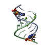
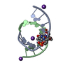
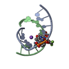
| Title | CRYSTAL STRUCTURE OF THE SELF-COMPLEMENTARY 5'-PURINE START DECAMER D(GCACGCGTGC) IN THE A-DNA CONFORMATION-PART II | ||||||||||||||||||
 Components Components | DNA (5'-D(* Keywords KeywordsDNA / A-DNA / DOUBLE HELIX | Function / homology | DNA |  Function and homology information Function and homology informationMethod |  X-RAY DIFFRACTION / Resolution: 1.9 Å X-RAY DIFFRACTION / Resolution: 1.9 Å  Authors AuthorsBan, C. / Sundaralingam, M. |  CitationJournal: Biophys.J. / Year: 1996 CitationJournal: Biophys.J. / Year: 1996Title: Crystal structure of the self-complementary 5'-purine start decamer d(GCACGCGTGC) in the A-DNA conformation. II. Authors: Ban, C. / Sundaralingam, M. History |
|
- Structure visualization
| Structure viewer | Molecule: MolmilJmol/JSmol |
|---|
- Downloads & links
-Download
| PDBx/mmCIF format | 260d.cif.gz | 15.2 KB | Display | PDBx/mmCIF format |
|---|---|---|---|---|
| PDB format | pdb260d.ent.gz | 9 KB | Display | PDB format |
| PDBx/mmJSON format | 260d.json.gz | Tree view | PDBx/mmJSON format | |
| Others |  Other downloads Other downloads |
-Validation report
| Summary document | 260d_validation.pdf.gz | 360.1 KB | Display | wwPDB validaton report |
|---|---|---|---|---|
| Full document | 260d_full_validation.pdf.gz | 362.6 KB | Display | |
| Data in XML | 260d_validation.xml.gz | 3.1 KB | Display | |
| Data in CIF | 260d_validation.cif.gz | 3.8 KB | Display | |
| Arichive directory | https://data.pdbj.org/pub/pdb/validation_reports/60/260dftp://data.pdbj.org/pub/pdb/validation_reports/60/260d | HTTPS FTP |
-Related structure data
| Similar structure data |
|---|


-Links
 PDBj
PDBj

- Assembly
| Deposited unit | 
| ||||||||
|---|---|---|---|---|---|---|---|---|---|
| 1 | 
| ||||||||
| Unit cell |
| ||||||||
| Components on special symmetry positions |
|
-Components
| #1: DNA chain | Mass: 3045.993 Da / Num. of mol.: 1 / Source method: isolated from a natural source |
|---|---|
| #2: Water | ChemComp-HOH /  Mass: 18.015 Da / Num. of mol.: 41 / Source method: isolated from a natural source / Formula: H2O Mass: 18.015 Da / Num. of mol.: 41 / Source method: isolated from a natural source / Formula: H2O |
-Experimental details
-Experiment
| Experiment | Method: X-RAY DIFFRACTION |
|---|
- Sample preparation
| Crystal | Density Matthews: 2.78 Å3/Da / Density % sol: 55.8 % | |||||||||||||||||||||||||||||||||||
|---|---|---|---|---|---|---|---|---|---|---|---|---|---|---|---|---|---|---|---|---|---|---|---|---|---|---|---|---|---|---|---|---|---|---|---|---|
| Crystal grow | Method: vapor diffusion / pH: 7 / Details: pH 7.00, VAPOR DIFFUSION / Temp details: ROOM TEMPERATURE | |||||||||||||||||||||||||||||||||||
| Components of the solutions |
| |||||||||||||||||||||||||||||||||||
| Crystal grow | *PLUS | |||||||||||||||||||||||||||||||||||
| Components of the solutions | *PLUS
|
-Data collection
| Diffraction | Ambient temp details: ROOM TEMPERATURE |
|---|---|
| Diffraction source | Source: ROTATING ANODE / Type: MACSCIENCE |
| Detector | Type: SIEMENS-NICOLET / Detector: AREA DETECTOR / Date: Dec 6, 1994 |
| Radiation | Monochromator: GRAPHITE / Monochromatic (M) / Laue (L): M / Scattering type: x-ray |
| Radiation wavelength | Relative weight: 1 |
| Reflection | Resolution: 1.9→8 Å / Num. all: 9346 / Num. obs: 2634 / % possible obs: 91 % / Observed criterion σ(F): 1 / Redundancy: 3.5 % / Rmerge(I) obs: 0.031 |
| Reflection | *PLUS Highest resolution: 1.9 Å / Lowest resolution: 8 Å / % possible obs: 91 % / Observed criterion σ(F): 1 / Redundancy: 3.5 % / Num. measured all: 9346 |
- Processing
| Software |
| ||||||||||||||||||||||||||||||||||||||||||||||||||||||||||||
|---|---|---|---|---|---|---|---|---|---|---|---|---|---|---|---|---|---|---|---|---|---|---|---|---|---|---|---|---|---|---|---|---|---|---|---|---|---|---|---|---|---|---|---|---|---|---|---|---|---|---|---|---|---|---|---|---|---|---|---|---|---|
| Refinement | Resolution: 1.9→8 Å / σ(F): 1 /
| ||||||||||||||||||||||||||||||||||||||||||||||||||||||||||||
| Refine Biso |
| ||||||||||||||||||||||||||||||||||||||||||||||||||||||||||||
| Refinement step | Cycle: LAST / Resolution: 1.9→8 Å
| ||||||||||||||||||||||||||||||||||||||||||||||||||||||||||||
| Refine LS restraints |
| ||||||||||||||||||||||||||||||||||||||||||||||||||||||||||||
| Xplor file | Serial no: 1 / Param file: PARAM_NDB.DNA | ||||||||||||||||||||||||||||||||||||||||||||||||||||||||||||
| Software | *PLUS Name: X-PLOR / Classification: refinement | ||||||||||||||||||||||||||||||||||||||||||||||||||||||||||||
| Refinement | *PLUS Highest resolution: 1.9 Å / Lowest resolution: 8 Å / σ(F): 1 | ||||||||||||||||||||||||||||||||||||||||||||||||||||||||||||
| Solvent computation | *PLUS | ||||||||||||||||||||||||||||||||||||||||||||||||||||||||||||
| Displacement parameters | *PLUS | ||||||||||||||||||||||||||||||||||||||||||||||||||||||||||||
| Refine LS restraints | *PLUS
|
