 Movie
Movie Controller


+ Open data

- Basic information
| Entry | Database: PDB / ID: 25qp | |||||||||||||||||||||
|---|---|---|---|---|---|---|---|---|---|---|---|---|---|---|---|---|---|---|---|---|---|---|
| Title | Cryo-EM Structure of PLPP3 | |||||||||||||||||||||
 Components Components | Phospholipid phosphatase 3 | |||||||||||||||||||||
 Keywords Keywords | MEMBRANE PROTEIN / Phosphatase | |||||||||||||||||||||
| Function / homology |  Function and homology information Function and homology informationsphingosine-1-phosphate phosphatase activity / ceramide-1-phosphate phosphatase activity / phosphatidate phosphatase / Sphingolipid catabolism / phosphatidate phosphatase activity / sphingosine metabolic process / positive regulation of endothelial cell-matrix adhesion / ceramide metabolic process / delta-catenin binding / phospholipid dephosphorylation ...sphingosine-1-phosphate phosphatase activity / ceramide-1-phosphate phosphatase activity / phosphatidate phosphatase / Sphingolipid catabolism / phosphatidate phosphatase activity / sphingosine metabolic process / positive regulation of endothelial cell-matrix adhesion / ceramide metabolic process / delta-catenin binding / phospholipid dephosphorylation / positive regulation of homotypic cell-cell adhesion / Hydrolases; Acting on ester bonds; Phosphoric-monoester hydrolases / sphingolipid catabolic process / cell-cell adhesion mediated by integrin / retrograde vesicle-mediated transport, Golgi to endoplasmic reticulum / phospholipid metabolic process / positive regulation of intracellular signal transduction / endoplasmic reticulum exit site / endoplasmic reticulum-Golgi intermediate compartment membrane / positive regulation of endothelial cell migration / integrin-mediated signaling pathway / adherens junction / trans-Golgi network / cell-cell adhesion / integrin binding / basolateral plasma membrane / protein stabilization / membrane raft / Golgi membrane / endoplasmic reticulum membrane / Golgi apparatus / signal transduction / positive regulation of transcription by RNA polymerase II / membrane / plasma membrane Similarity search - Function | |||||||||||||||||||||
| Biological species |  Homo sapiens (human) Homo sapiens (human) | |||||||||||||||||||||
| Method | ELECTRON MICROSCOPY / single particle reconstruction / cryo EM / Resolution: 2.9 Å | |||||||||||||||||||||
 Authors Authors | Long, T. / Wu, Y. | |||||||||||||||||||||
| Funding support | 1items
| |||||||||||||||||||||
 Citation Citation | Journal: Nat Commun / Year: 2026 Title: Structural basis of PLPP3-mediated lipid phosphate dephosphorylation and its role in melanoma. Authors: Yingjie Wu / Di Xiao / Xingfan Li / Keyu Wang / Hongxu Zhang / Yuanyuan Zhao / Di Wu / Ruxi Qi / Mi Zhou / Han Han / Tao Long /  Abstract: Lipid phosphates serve as signaling molecules involved in diverse cellular processes such as cell proliferation, migration, angiogenesis, inflammation, immunity and cancer progression. Phospholipid ...Lipid phosphates serve as signaling molecules involved in diverse cellular processes such as cell proliferation, migration, angiogenesis, inflammation, immunity and cancer progression. Phospholipid phosphatases (PLPPs) modulate these signals by catalyzing the dephosphorylation of lipid phosphates. Here, we report the cryo-EM structure of PLPP3, revealing a tetrameric assembly. PLPP3 contains six transmembrane helices (TMs) and an extracellular domain that contains two extracellular loops. TMs 1-4 create a hydrophobic cleft that holds the tails of a phospholipid while the extracellular domain forms a positively charged pocket to accommodate the polar head group. Two conserved catalytic histidine residues in this pocket coordinate a putative zinc ion previously identified as a PLPP3 inhibitor. Structural mapping of somatic mutations with functional analysis reveals that PLPP3 acts as a tumor suppressor in melanoma. Together, our findings provide critical insights into the structure, substrate engagement, inhibitory mechanism, and cancer-related function of PLPP3. | |||||||||||||||||||||
| History |
|
- Structure visualization
| Structure viewer | Molecule: MolmilJmol/JSmol |
|---|
- Downloads & links
-Download
| PDBx/mmCIF format | 25qp.cif.gz | 218.1 KB | Display | PDBx/mmCIF format |
|---|---|---|---|---|
| PDB format | pdb25qp.ent.gz | 174 KB | Display | PDB format |
| PDBx/mmJSON format | 25qp.json.gz | Tree view | PDBx/mmJSON format | |
| Others |  Other downloads Other downloads |
-Validation report
| Arichive directory | https://data.pdbj.org/pub/pdb/validation_reports/5q/25qpftp://data.pdbj.org/pub/pdb/validation_reports/5q/25qp | HTTPS FTP |
|---|
-Related structure data
| Related structure data |  80306MC M: map data used to model this data C: citing same article ( |
|---|---|
| Similar structure data |

-Links
 PDBj
PDBj- Assembly
| Deposited unit | 
|
|---|---|
| 1 |
|
-Components
-Protein / Sugars , 2 types, 8 molecules ABCD
| #1: Protein | Mass: 36024.758 Da / Num. of mol.: 4 Source method: isolated from a genetically manipulated source Source: (gene. exp.) Homo sapiens (human) / Gene: PLPP3, LPP3, PPAP2B / Production host: Homo sapiens (human)References: UniProt: O14495, Hydrolases; Acting on ester bonds; Phosphoric-monoester hydrolases, phosphatidate phosphatase #5: Sugar | ChemComp-NAG /  Type: D-saccharide, beta linking / Mass: 221.208 Da / Num. of mol.: 4 / Source method: obtained synthetically / Formula: C8H15NO6 Type: D-saccharide, beta linking / Mass: 221.208 Da / Num. of mol.: 4 / Source method: obtained synthetically / Formula: C8H15NO6 |
|---|
-Non-polymers , 4 types, 16 molecules 



| #2: Chemical | ChemComp-LBN /  Mass: 760.076 Da / Num. of mol.: 4 / Source method: obtained synthetically / Formula: C42H82NO8P / Feature type: SUBJECT OF INVESTIGATION / Comment: phospholipid*YM Mass: 760.076 Da / Num. of mol.: 4 / Source method: obtained synthetically / Formula: C42H82NO8P / Feature type: SUBJECT OF INVESTIGATION / Comment: phospholipid*YM#3: Chemical | ChemComp-PX2 / 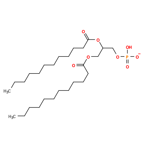 Mass: 535.671 Da / Num. of mol.: 4 / Source method: isolated from a natural source / Formula: C27H52O8P / Feature type: SUBJECT OF INVESTIGATION Mass: 535.671 Da / Num. of mol.: 4 / Source method: isolated from a natural source / Formula: C27H52O8P / Feature type: SUBJECT OF INVESTIGATION#4: Chemical | ChemComp-ZN /  Mass: 65.409 Da / Num. of mol.: 4 / Source method: obtained synthetically / Formula: Zn Mass: 65.409 Da / Num. of mol.: 4 / Source method: obtained synthetically / Formula: Zn#6: Water | ChemComp-HOH / | Mass: 18.015 Da / Num. of mol.: 4 / Source method: isolated from a natural source / Formula: H2O |
|---|
-Details
| Has ligand of interest | Y |
|---|---|
| Has protein modification | Y |
-Experimental details
-Experiment
| Experiment | Method: ELECTRON MICROSCOPY |
|---|---|
| EM experiment | Aggregation state: PARTICLE / 3D reconstruction method: single particle reconstruction |
- Sample preparation
| Component | Name: PLPP3 / Type: CELL / Entity ID: #1 / Source: RECOMBINANT |
|---|---|
| Source (natural) | Organism: Homo sapiens (human) |
| Source (recombinant) | Organism: Homo sapiens (human) |
| Buffer solution | pH: 7.5 |
| Specimen | Embedding applied: NO / Shadowing applied: NO / Staining applied: NO / Vitrification applied: YES |
| Vitrification | Cryogen name: ETHANE |
- Electron microscopy imaging
| Microscopy | Model: FEI MORGAGNI |
|---|---|
| Electron gun | Electron source:  FIELD EMISSION GUN / Accelerating voltage: 300 kV / Illumination mode: FLOOD BEAM FIELD EMISSION GUN / Accelerating voltage: 300 kV / Illumination mode: FLOOD BEAM |
| Electron lens | Mode: DARK FIELD / Nominal defocus max: 1800 nm / Nominal defocus min: 1200 nm |
| Image recording | Electron dose: 50 e/Å2 / Film or detector model: GATAN K3 (6k x 4k) |
- Processing
| EM software |
| ||||||||||||||||
|---|---|---|---|---|---|---|---|---|---|---|---|---|---|---|---|---|---|
| CTF correction | Type: PHASE FLIPPING AND AMPLITUDE CORRECTION | ||||||||||||||||
| 3D reconstruction | Resolution: 2.9 Å / Resolution method: FSC 0.143 CUT-OFF / Num. of particles: 289649 / Symmetry type: POINT |
