[English] 日本語
 Yorodumi
- PDB-242d: MAD PHASING STRATEGIES EXPLORED WITH A BROMINATED OLIGONUCLEOTIDE... -
+ Open data
- Basic information
| Entry | Database: PDB / ID: 242d | ||||||||||||||||||
|---|---|---|---|---|---|---|---|---|---|---|---|---|---|---|---|---|---|---|---|
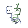
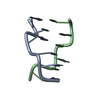
| Title | MAD PHASING STRATEGIES EXPLORED WITH A BROMINATED OLIGONUCLEOTIDE CRYSTAL AT 1.65 A RESOLUTION. | ||||||||||||||||||
 Components Components | DNA (5'-D(* Keywords KeywordsDNA / Z-DNA / DOUBLE HELIX / MODIFIED | Function / homology | DNA |  Function and homology information Function and homology informationMethod |  X-RAY DIFFRACTION / X-RAY DIFFRACTION /  SYNCHROTRON / SYNCHROTRON /  MAD / Resolution: 1.65 Å MAD / Resolution: 1.65 Å  Authors AuthorsPeterson, M.R. / Harrop, S.J. / McSweeney, S.M. / Leonard, G.A. / Thompson, A.W. / Hunter, W.N. / Helliwell, J.R. |  Citation Citation Journal: J.Synchrotron Radiat. / Year: 1996 Journal: J.Synchrotron Radiat. / Year: 1996Title: MAD Phasing Strategies Explored with a Brominated Oligonucleotide Crystal at 1.65A Resolution. Authors: Peterson, M.R. / Harrop, S.J. / McSweeney, S.M. / Leonard, G.A. / Thompson, A.W. / Hunter, W.N. / Helliwell, J.R. #1:  Journal: Nature / Year: 1979 Journal: Nature / Year: 1979Title: Molecular Structure of a Left-Handed Double Helical DNA Fragment at Atomic Resolution Authors: Wang, A.H.-J. / Quigley, G.J. / Kolpak, F.J. / Crawford, J.L. / Van Boom, J.H. / Van Der Marel, G. / Rich, A. History |
|
- Structure visualization
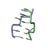
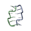
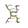
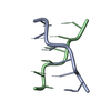
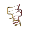
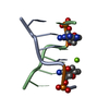
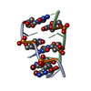
| Structure viewer | Molecule:  Molmil Molmil Jmol/JSmol Jmol/JSmol |
|---|
- Downloads & links
- Download
| PDBx/mmCIF format |  242d.cif.gz 242d.cif.gz | 16 KB | Display |  PDBx/mmCIF format PDBx/mmCIF format |
|---|---|---|---|---|
| PDB format |  pdb242d.ent.gz pdb242d.ent.gz | 10.4 KB | Display |  PDB format PDB format |
| PDBx/mmJSON format |  242d.json.gz 242d.json.gz | Tree view |  PDBx/mmJSON format PDBx/mmJSON format | |
| Others |  Other downloads Other downloads |
-Validation report
| Summary document |  242d_validation.pdf.gz 242d_validation.pdf.gz | 371 KB | Display |  wwPDB validaton report wwPDB validaton report |
|---|---|---|---|---|
| Full document |  242d_full_validation.pdf.gz 242d_full_validation.pdf.gz | 373.5 KB | Display | |
| Data in XML |  242d_validation.xml.gz 242d_validation.xml.gz | 3.8 KB | Display | |
| Data in CIF |  242d_validation.cif.gz 242d_validation.cif.gz | 4.8 KB | Display | |
| Arichive directory |  https://data.pdbj.org/pub/pdb/validation_reports/42/242d https://data.pdbj.org/pub/pdb/validation_reports/42/242d ftp://data.pdbj.org/pub/pdb/validation_reports/42/242d ftp://data.pdbj.org/pub/pdb/validation_reports/42/242d | HTTPS FTP |
-Related structure data
| Similar structure data |
|---|
- Links
Links
- Assembly
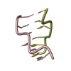
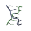
| Deposited unit | 
| ||||||||
|---|---|---|---|---|---|---|---|---|---|
| 1 |
| ||||||||
| Unit cell |
|
- Components
| #1: DNA chain | Mass: 1889.101 Da / Num. of mol.: 2 / Source method: obtained synthetically #2: Water | ChemComp-HOH / | |
|---|
-Experimental details
-Experiment
| Experiment | Method:  X-RAY DIFFRACTION X-RAY DIFFRACTION |
|---|
- Sample preparation
| Crystal | Density Matthews: 1.65 Å3/Da / Density % sol: 25.55 % |
|---|---|
| Crystal grow | *PLUS Method: unknown |
-Data collection
| Diffraction source | Source:  SYNCHROTRON / Site: SYNCHROTRON / Site:  SRS SRS  / Beamline: PX9.5 / Beamline: PX9.5 |
|---|---|
| Detector | Type: MARRESEARCH / Detector: IMAGE PLATE / Date: Sep 15, 1993 |
| Radiation | Monochromatic (M) / Laue (L): M / Scattering type: x-ray |
| Radiation wavelength | Relative weight: 1 |
- Processing
| Software |
| ||||||||||||||||||||||||||||||||||||||||||||||||||||||||||||||||||||||||||||||||||||
|---|---|---|---|---|---|---|---|---|---|---|---|---|---|---|---|---|---|---|---|---|---|---|---|---|---|---|---|---|---|---|---|---|---|---|---|---|---|---|---|---|---|---|---|---|---|---|---|---|---|---|---|---|---|---|---|---|---|---|---|---|---|---|---|---|---|---|---|---|---|---|---|---|---|---|---|---|---|---|---|---|---|---|---|---|---|
| Refinement | Method to determine structure:  MAD / Resolution: 1.65→8 Å / σ(F): 0 / MAD / Resolution: 1.65→8 Å / σ(F): 0 /
| ||||||||||||||||||||||||||||||||||||||||||||||||||||||||||||||||||||||||||||||||||||
| Refine Biso |
| ||||||||||||||||||||||||||||||||||||||||||||||||||||||||||||||||||||||||||||||||||||
| Refinement step | Cycle: LAST / Resolution: 1.65→8 Å
| ||||||||||||||||||||||||||||||||||||||||||||||||||||||||||||||||||||||||||||||||||||
| Refine LS restraints |
| ||||||||||||||||||||||||||||||||||||||||||||||||||||||||||||||||||||||||||||||||||||
| Software | *PLUS Name: PROLSQ / Classification: refinement | ||||||||||||||||||||||||||||||||||||||||||||||||||||||||||||||||||||||||||||||||||||
| Refine LS restraints | *PLUS
|
 Movie
Movie Controller








 PDBj
PDBj


