Movie
 Controller


+ Open data

- Basic information
| Entry | Database: PDB / ID: 22vd | ||||||||||||
|---|---|---|---|---|---|---|---|---|---|---|---|---|---|
| Title | Cryo-EM structure of DOCK11 | ||||||||||||
 Components | Dedicator of cytokinesis protein 11 | ||||||||||||
 Keywords | SIGNALING PROTEIN / RhoGEF | ||||||||||||
| Function / homology |  Function and homology information marginal zone B cell differentiation / B cell homeostasis / regulation of Rho protein signal transduction / positive regulation of filopodium assembly / small GTPase-mediated signal transduction / CDC42 GTPase cycle / positive regulation of GTPase activity / RAC1 GTPase cycle / guanyl-nucleotide exchange factor activity / small GTPase binding ...marginal zone B cell differentiation / B cell homeostasis / regulation of Rho protein signal transduction / positive regulation of filopodium assembly / small GTPase-mediated signal transduction / CDC42 GTPase cycle / positive regulation of GTPase activity / RAC1 GTPase cycle / guanyl-nucleotide exchange factor activity / small GTPase binding / Factors involved in megakaryocyte development and platelet production / cytosol Similarity search - Function | ||||||||||||
| Biological species |  Homo sapiens (human) | ||||||||||||
| Method | ELECTRON MICROSCOPY / single particle reconstruction / cryo EM / Resolution: 3.8 Å | ||||||||||||
 Authors | Hisano, T. / Kukimoto-Niino, M. / Shinoda, T. / Katsura, K. / Shirouzu, M. | ||||||||||||
| Funding support |  Japan, 3items
| ||||||||||||
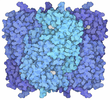
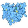
 Citation | Journal: Proc Natl Acad Sci U S A / Year: 2026 Title: Structural basis for membrane-dependent positive feedback regulation of DOCK11 by activated Cdc42. Authors: Tamao Hisano / Takehiro Shinoda / Mutsuko Kukimoto-Niino / Kazushige Katsura / Ryohei Kondo / Yoshiko Ishizuka-Katsura / Yukako Miyata-Yabuki / Kazuharu Hanada / Reiko Nakagawa / Akihiko ...Authors: Tamao Hisano / Takehiro Shinoda / Mutsuko Kukimoto-Niino / Kazushige Katsura / Ryohei Kondo / Yoshiko Ishizuka-Katsura / Yukako Miyata-Yabuki / Kazuharu Hanada / Reiko Nakagawa / Akihiko Nishikimi / Mikako Shirouzu / Abstract: The guanine nucleotide exchange factor DOCK11 is widely expressed across tissues and activates Cdc42, a Rho family GTPase that regulates cytoskeletal remodeling and cell polarity. Loss-of-function ...The guanine nucleotide exchange factor DOCK11 is widely expressed across tissues and activates Cdc42, a Rho family GTPase that regulates cytoskeletal remodeling and cell polarity. Loss-of-function mutations in DOCK11 have recently been linked to inborn errors of immunity. DOCK11 has also been implicated in facilitating hepatitis B virus infection in hepatocytes and has emerged as a potential target for therapeutic interventions. A positive feedback mechanism has been proposed for DOCK11-mediated Cdc42 activation; however, the structural basis for this regulation has remained unclear. Here we report the cryoelectron microscopy structures of full-length DOCK11 as an apo dimer, a binary complex with nucleotide-free Cdc42, and a ternary complex containing both nucleotide-free and activated Cdc42. DOCK11 predominantly adopts a U-shaped dimer that binds to nucleotide-free Cdc42 via the catalytic domain, whereas activated Cdc42 engages a distal site within the armadillo-repeat domain. Biochemical and cellular analyses demonstrate that binding of activated Cdc42 is critical for enhancing the membrane-dependent nucleotide exchange activity of DOCK11, promoting its localization to the plasma membrane, and facilitating intracellular activation of Cdc42. We also determined the structure of the membrane-bound ternary complex using membrane-coated grids. Strikingly, the ternary complex adopts an extended conformation that aligns multiple membrane-contact elements, revealing how activated Cdc42 stabilizes DOCK11 membrane binding for efficient exchange activity. Collectively, these findings provide a structural framework for the coordinated mechanism underlying membrane-dependent positive feedback in Rho GTPase signaling. | ||||||||||||
| History |
|
- Structure visualization
| Structure viewer | Molecule: MolmilJmol/JSmol |
|---|
- Downloads & links
-Download
| PDBx/mmCIF format | 22vd.cif.gz | 1.1 MB | Display | PDBx/mmCIF format |
|---|---|---|---|---|
| PDB format | pdb22vd.ent.gz | 974.6 KB | Display | PDB format |
| PDBx/mmJSON format | 22vd.json.gz | Tree view | PDBx/mmJSON format | |
| Others |  Other downloads |
-Validation report
| Arichive directory | https://data.pdbj.org/pub/pdb/validation_reports/2v/22vdftp://data.pdbj.org/pub/pdb/validation_reports/2v/22vd | HTTPS FTP |
|---|
-Related structure data
| Related structure data |  68711MC  22veC  22vfC  22vgC M: map data used to model this data C: citing same article ( |
|---|---|
| Similar structure data |


-Links
 PDBj
- Assembly
| Deposited unit | 
|
|---|---|
| 1 |
|
-Components
| #1: Protein | Mass: 238396.047 Da / Num. of mol.: 2 Source method: isolated from a genetically manipulated source Source: (gene. exp.) Homo sapiens (human) / Gene: DOCK11, ZIZ2 / Cell line (production host): FreeStyle 293-F / Production host: Homo sapiens (human) / References: UniProt: Q5JSL3Has protein modification | N | |
|---|
-Experimental details
-Experiment
| Experiment | Method: ELECTRON MICROSCOPY |
|---|---|
| EM experiment | Aggregation state: PARTICLE / 3D reconstruction method: single particle reconstruction |
- Sample preparation
| Component | Name: The DOCK11 dimer / Type: ORGANELLE OR CELLULAR COMPONENT / Entity ID: all / Source: RECOMBINANT |
|---|---|
| Molecular weight | Experimental value: NO |
| Source (natural) | Organism: Homo sapiens (human) |
| Source (recombinant) | Organism: Homo sapiens (human) |
| Buffer solution | pH: 8 |
| Specimen | Embedding applied: NO / Shadowing applied: NO / Staining applied: NO / Vitrification applied: YES |
| Vitrification | Instrument: FEI VITROBOT MARK IV / Cryogen name: ETHANE |
- Electron microscopy imaging
| Experimental equipment |  Model: Titan Krios / Image courtesy: FEI Company |
|---|---|
| Microscopy | Model: TFS KRIOS |
| Electron gun | Electron source:  FIELD EMISSION GUN / Accelerating voltage: 300 kV / Illumination mode: FLOOD BEAM |
| Electron lens | Mode: BRIGHT FIELD / Nominal defocus max: 2200 nm / Nominal defocus min: 800 nm |
| Specimen holder | Cryogen: NITROGEN / Specimen holder model: FEI TITAN KRIOS AUTOGRID HOLDER |
| Image recording | Electron dose: 50.3 e/Å2 / Film or detector model: GATAN K3 BIOCONTINUUM (6k x 4k) |
- Processing
| EM software |
| ||||||||||||||||||||||||||||||||||||
|---|---|---|---|---|---|---|---|---|---|---|---|---|---|---|---|---|---|---|---|---|---|---|---|---|---|---|---|---|---|---|---|---|---|---|---|---|---|
| CTF correction | Type: PHASE FLIPPING AND AMPLITUDE CORRECTION | ||||||||||||||||||||||||||||||||||||
| Symmetry | Point symmetry: C2 (2 fold cyclic) | ||||||||||||||||||||||||||||||||||||
| 3D reconstruction | Resolution: 3.8 Å / Resolution method: FSC 0.143 CUT-OFF / Num. of particles: 511163 / Symmetry type: POINT | ||||||||||||||||||||||||||||||||||||
| Atomic model building | Protocol: RIGID BODY FIT / Space: REAL | ||||||||||||||||||||||||||||||||||||
| Atomic model building | PDB-ID: 22VF Pdb chain-ID: A / Accession code: 22VF / Details: DOCK11 / Source name: PDB / Type: experimental model | ||||||||||||||||||||||||||||||||||||
| Refinement | Highest resolution: 3.8 Å Stereochemistry target values: REAL-SPACE (WEIGHTED MAP SUM AT ATOM CENTERS) | ||||||||||||||||||||||||||||||||||||
| Refine LS restraints |
|