 Movie
Movie Controller


+ Open data

- Basic information
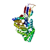
| Entry | Database: PDB / ID: 22gt | ||||||
|---|---|---|---|---|---|---|---|
| Title | a novel GH8 family xylanase BiXyn8A | ||||||
 Components Components | cellulase | ||||||
 Keywords Keywords | HYDROLASE / GH8 family / endoxylanase | ||||||
| Function / homology | Glycoside hydrolase, family 8 / Glycosyl hydrolases family 8 / cellulase / cellulase activity / Six-hairpin glycosidase-like superfamily / Six-hairpin glycosidase superfamily / cellulose catabolic process / cellulase Function and homology information Function and homology information | ||||||
| Biological species |  Bacteroides intestinalis DSM 17393 (bacteria) Bacteroides intestinalis DSM 17393 (bacteria) | ||||||
| Method |  X-RAY DIFFRACTION / MOLECULAR REPLACEMENT / Resolution: 1.80000297631 Å X-RAY DIFFRACTION / MOLECULAR REPLACEMENT / Resolution: 1.80000297631 Å | ||||||
 Authors Authors | Wei, X. / Yun, L. | ||||||
| Funding support |  China, 1items China, 1items
| ||||||
 Citation Citation | Journal: Bioresour Technol / Year: 2026 Title: Biochemical and structural characterization of a novel glycoside hydrolase family 8 endoxylanase with broad-spectrum xylooligosaccharide production. Authors: Liu, Y. / Xie, W. / Zhang, Y. / Liang, S. / Wang, S. / Zhan, R. / Wang, C. / Wang, K. | ||||||
| History |
|
- Structure visualization
| Structure viewer | Molecule: MolmilJmol/JSmol |
|---|
- Downloads & links
-Download
| PDBx/mmCIF format | 22gt.cif.gz | 212.3 KB | Display | PDBx/mmCIF format |
|---|---|---|---|---|
| PDB format | pdb22gt.ent.gz | 135.2 KB | Display | PDB format |
| PDBx/mmJSON format | 22gt.json.gz | Tree view | PDBx/mmJSON format | |
| Others |  Other downloads Other downloads |
-Validation report
| Arichive directory | https://data.pdbj.org/pub/pdb/validation_reports/2g/22gtftp://data.pdbj.org/pub/pdb/validation_reports/2g/22gt | HTTPS FTP |
|---|
-Related structure data

-Links
 PDBj
PDBj
- Assembly
| Deposited unit | 
| ||||||||||||
|---|---|---|---|---|---|---|---|---|---|---|---|---|---|
| 1 | 
| ||||||||||||
| 2 | 
| ||||||||||||
| Unit cell |
|
-Components
| #1: Protein | Mass: 47249.617 Da / Num. of mol.: 2 Source method: isolated from a genetically manipulated source Source: (gene. exp.) Bacteroides intestinalis DSM 17393 (bacteria)Gene: BACINT_04210 / Production host: #2: Water | ChemComp-HOH / |  Mass: 18.015 Da / Num. of mol.: 380 / Source method: isolated from a natural source / Formula: H2O Mass: 18.015 Da / Num. of mol.: 380 / Source method: isolated from a natural source / Formula: H2OHas protein modification | N | |
|---|
-Experimental details
-Experiment
| Experiment | Method: X-RAY DIFFRACTION / Number of used crystals: 1 |
|---|
- Sample preparation
| Crystal | Density Matthews: 2.22 Å3/Da / Density % sol: 44.68 % |
|---|---|
| Crystal grow | Temperature: 298 K / Method: vapor diffusion, sitting drop / Details: PEG3350, Sodium Malonate |
-Data collection
| Diffraction | Mean temperature: 100 K / Serial crystal experiment: N |
|---|---|
| Diffraction source | Source: ROTATING ANODE / Type: RIGAKU / Wavelength: 1.54 Å |
| Detector | Type: MAR CCD 130 mm / Detector: CCD / Date: Nov 10, 2020 |
| Radiation | Protocol: SINGLE WAVELENGTH / Monochromatic (M) / Laue (L): M / Scattering type: x-ray |
| Radiation wavelength | Wavelength: 1.54 Å / Relative weight: 1 |
| Reflection | Resolution: 1.8→25.01 Å / Num. obs: 386257 / % possible obs: 98.98 % / Redundancy: 4.8 % / Biso Wilson estimate: 19.7758750919 Å2 / CC1/2: 0.997 / CC star: 0.999 / Net I/σ(I): 14.88 |
| Reflection shell | Resolution: 1.8→10 Å / Mean I/σ(I) obs: 14.88 / Num. unique obs: 77988 / CC1/2: 0.997 |
- Processing
| Software |
| |||||||||||||||||||||||||||||||||||||||||||||||||||||||||||||||||||||||||||||||||||||||||||||||||||||||||||||||||||||||||||||||||||||||||||||||||||||||||||||||||||||||||||||||||||||||||||||||||||||||||||
|---|---|---|---|---|---|---|---|---|---|---|---|---|---|---|---|---|---|---|---|---|---|---|---|---|---|---|---|---|---|---|---|---|---|---|---|---|---|---|---|---|---|---|---|---|---|---|---|---|---|---|---|---|---|---|---|---|---|---|---|---|---|---|---|---|---|---|---|---|---|---|---|---|---|---|---|---|---|---|---|---|---|---|---|---|---|---|---|---|---|---|---|---|---|---|---|---|---|---|---|---|---|---|---|---|---|---|---|---|---|---|---|---|---|---|---|---|---|---|---|---|---|---|---|---|---|---|---|---|---|---|---|---|---|---|---|---|---|---|---|---|---|---|---|---|---|---|---|---|---|---|---|---|---|---|---|---|---|---|---|---|---|---|---|---|---|---|---|---|---|---|---|---|---|---|---|---|---|---|---|---|---|---|---|---|---|---|---|---|---|---|---|---|---|---|---|---|---|---|---|---|---|---|---|---|
| Refinement | Method to determine structure: MOLECULAR REPLACEMENT / Resolution: 1.80000297631→25.0082581824 Å / SU ML: 0.192869828307 / Cross valid method: FREE R-VALUE / σ(F): 1.3493385646 / Phase error: 21.6974524398 Stereochemistry target values: GeoStd + Monomer Library + CDL v1.2
| |||||||||||||||||||||||||||||||||||||||||||||||||||||||||||||||||||||||||||||||||||||||||||||||||||||||||||||||||||||||||||||||||||||||||||||||||||||||||||||||||||||||||||||||||||||||||||||||||||||||||||
| Solvent computation | Shrinkage radii: 0.9 Å / VDW probe radii: 1.11 Å / Solvent model: FLAT BULK SOLVENT MODEL | |||||||||||||||||||||||||||||||||||||||||||||||||||||||||||||||||||||||||||||||||||||||||||||||||||||||||||||||||||||||||||||||||||||||||||||||||||||||||||||||||||||||||||||||||||||||||||||||||||||||||||
| Displacement parameters | Biso mean: 22.2505307492 Å2 | |||||||||||||||||||||||||||||||||||||||||||||||||||||||||||||||||||||||||||||||||||||||||||||||||||||||||||||||||||||||||||||||||||||||||||||||||||||||||||||||||||||||||||||||||||||||||||||||||||||||||||
| Refinement step | Cycle: LAST / Resolution: 1.80000297631→25.0082581824 Å
| |||||||||||||||||||||||||||||||||||||||||||||||||||||||||||||||||||||||||||||||||||||||||||||||||||||||||||||||||||||||||||||||||||||||||||||||||||||||||||||||||||||||||||||||||||||||||||||||||||||||||||
| Refine LS restraints |
| |||||||||||||||||||||||||||||||||||||||||||||||||||||||||||||||||||||||||||||||||||||||||||||||||||||||||||||||||||||||||||||||||||||||||||||||||||||||||||||||||||||||||||||||||||||||||||||||||||||||||||
| LS refinement shell |
|
