Mass: 9650.206 Da / Num. of mol.: 1 Source method: isolated from a genetically manipulated source Source: (gene. exp.) Homo sapiens (human) / Gene: SNN / Plasmid: pMal-c2x / Species (production host): Escherichia coli / Production host: Escherichia coli BL21(DE3) (bacteria) / Strain (production host): BL21(DE3) / References: UniProt: O75324
-
Experimental details
-
Experiment
Experiment
Method: SOLUTION NMR
NMR experiment
Type: 3D 15N-separated NOESY
NMR details
Text: Backbone assignments were made using 3D-TROSY based HNCA, HN(CO)CA, HNCACB, CBCA(CO)NH and HNCO experiments. Side chains and NOEs were assigned with 3D [1H, 15N]-TOCSY-HSQC and 3D [1H,15N]-NOESY-HSQC experiments
-
Sample preparation
Details
Contents: 0.5-0.7 mM Stannin U-15N or U-15N/13C; 20 mM phosphate buffer; 600 mM d25-sodium dodecyl sulfate; 90% H2O; 10% D2O; 0.02% sodium azide; 0.7 mM DTT Solvent system: 90% H2O/10% D2O
Sample conditions
Ionic strength: 120 mM NaCl / pH: 4 / Pressure: ambient / Temperature: 333 K
-
NMR measurement
Radiation
Protocol: SINGLE WAVELENGTH / Monochromatic (M) / Laue (L): M
In the structure databanks used in Yorodumi, some data are registered as the other names, "COVID-19 virus" and "2019-nCoV". Here are the details of the virus and the list of structure data.
Jan 31, 2019. EMDB accession codes are about to change! (news from PDBe EMDB page)
EMDB accession codes are about to change! (news from PDBe EMDB page)
The allocation of 4 digits for EMDB accession codes will soon come to an end. Whilst these codes will remain in use, new EMDB accession codes will include an additional digit and will expand incrementally as the available range of codes is exhausted. The current 4-digit format prefixed with “EMD-” (i.e. EMD-XXXX) will advance to a 5-digit format (i.e. EMD-XXXXX), and so on. It is currently estimated that the 4-digit codes will be depleted around Spring 2019, at which point the 5-digit format will come into force.
The EM Navigator/Yorodumi systems omit the EMD- prefix.
Related info.:Q: What is EMD? / ID/Accession-code notation in Yorodumi/EM Navigator
Yorodumi is a browser for structure data from EMDB, PDB, SASBDB, etc.
This page is also the successor to EM Navigator detail page, and also detail information page/front-end page for Omokage search.
The word "yorodu" (or yorozu) is an old Japanese word meaning "ten thousand". "mi" (miru) is to see.
Related info.:EMDB / PDB / SASBDB / Comparison of 3 databanks / Yorodumi Search / Aug 31, 2016. New EM Navigator & Yorodumi / Yorodumi Papers / Jmol/JSmol / Function and homology information / Changes in new EM Navigator and Yorodumi
 Movie
Movie Controller


 Open data
 Basic information
 Components
 Keywords
 Function and homology information
 Homo sapiens (human)
 Authors
 Citation
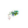
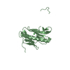
 Structure visualization
 Downloads & links
 Other downloads
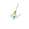


 PDBj
PDBj Assembly

 Sample preparation
 Processing
 NMRPipe