 Movie
Movie Controller

[English] 日本語
 Yorodumi
- PDB-1zd4: Human soluble epoxide hydrolase 4-(3-cyclohexyluriedo)-hexanoic a... -
+ Open data

- Basic information
| Entry | Database: PDB / ID: 1zd4 | ||||||
|---|---|---|---|---|---|---|---|
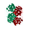
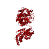
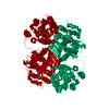
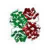
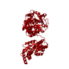
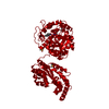
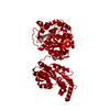
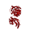
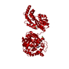
| Title | Human soluble epoxide hydrolase 4-(3-cyclohexyluriedo)-hexanoic acid complex | ||||||
 Components Components | epoxide hydrolase 2, cytoplasmic | ||||||
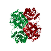
 Keywords Keywords | HYDROLASE / domain swapped dimer | ||||||
| Function / homology |  Function and homology information Function and homology informationlipid-phosphate phosphatase / 10-hydroxy-9-(phosphonooxy)octadecanoate phosphatase activity / stilbene catabolic process / Biosynthesis of maresins / epoxide metabolic process / phospholipid dephosphorylation / lipid phosphatase activity / soluble epoxide hydrolase / Synthesis of epoxy (EET) and dihydroxyeicosatrienoic acids (DHET) / lysophosphatidic acid phosphatase activity ...lipid-phosphate phosphatase / 10-hydroxy-9-(phosphonooxy)octadecanoate phosphatase activity / stilbene catabolic process / Biosynthesis of maresins / epoxide metabolic process / phospholipid dephosphorylation / lipid phosphatase activity / soluble epoxide hydrolase / Synthesis of epoxy (EET) and dihydroxyeicosatrienoic acids (DHET) / lysophosphatidic acid phosphatase activity / epoxide hydrolase activity / dephosphorylation / regulation of cholesterol metabolic process / peroxisomal matrix / phosphatase activity / toxic substance binding / cholesterol homeostasis / Peroxisomal protein import / regulation of cell growth / response to toxic substance / peroxisome / positive regulation of gene expression / magnesium ion binding / protein homodimerization activity / extracellular exosome / cytosol Similarity search - Function | ||||||
| Biological species |  Homo sapiens (human) Homo sapiens (human) | ||||||
| Method |  X-RAY DIFFRACTION / MOLECULAR REPLACEMENT / Resolution: 2.7 Å X-RAY DIFFRACTION / MOLECULAR REPLACEMENT / Resolution: 2.7 Å | ||||||
 Authors Authors | Gomez, G.A. / Morisseau, C. / Hammock, B.D. / Christianson, D.W. | ||||||
 Citation Citation | Journal: Protein Sci. / Year: 2006 Title: Human soluble epoxide hydrolase: structural basis of inhibition by 4-(3-cyclohexylureido)-carboxylic acids Authors: Gomez, G.A. / Morisseau, C. / Hammock, B.D. / Christianson, D.W. | ||||||
| History |
|
- Structure visualization
| Structure viewer | Molecule: MolmilJmol/JSmol |
|---|
- Downloads & links
-Download
| PDBx/mmCIF format | 1zd4.cif.gz | 113 KB | Display | PDBx/mmCIF format |
|---|---|---|---|---|
| PDB format | pdb1zd4.ent.gz | 87.8 KB | Display | PDB format |
| PDBx/mmJSON format | 1zd4.json.gz | Tree view | PDBx/mmJSON format | |
| Others |  Other downloads Other downloads |
-Validation report
| Arichive directory | https://data.pdbj.org/pub/pdb/validation_reports/zd/1zd4ftp://data.pdbj.org/pub/pdb/validation_reports/zd/1zd4 | HTTPS FTP |
|---|
-Related structure data
| Related structure data | 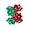 1zd2C 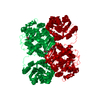 1zd3C  1zd5C 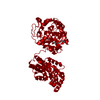 1s8oS S: Starting model for refinement C: citing same article ( |
|---|---|
| Similar structure data |


-Links
 PDBj
PDBj


- Assembly
| Deposited unit | 
| ||||||||
|---|---|---|---|---|---|---|---|---|---|
| 1 | 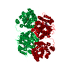
| ||||||||
| Unit cell |
| ||||||||
| Components on special symmetry positions |
| ||||||||
| Details | The second part of the biological assembly is generated by the symmetry_op=(-Y,-X,-Z+1/6) dx= 1 dy= 1 dz= 1 distance= 0.001 |
-Components
| #1: Protein | Mass: 62685.617 Da / Num. of mol.: 1 Source method: isolated from a genetically manipulated source Source: (gene. exp.) Homo sapiens (human) / Gene: EPHX2 / Plasmid: ACHSEH1 / Cell line (production host): SF9 / Production host:   Spodoptera frugiperda (fall armyworm) / References: UniProt: P34913, epoxide hydrolase Spodoptera frugiperda (fall armyworm) / References: UniProt: P34913, epoxide hydrolase |
|---|---|
| #2: Chemical | ChemComp-MG /   Mass: 24.305 Da / Num. of mol.: 1 / Source method: obtained synthetically / Formula: Mg Mass: 24.305 Da / Num. of mol.: 1 / Source method: obtained synthetically / Formula: Mg |
| #3: Chemical | ChemComp-PO4 /   Mass: 94.971 Da / Num. of mol.: 1 / Source method: obtained synthetically / Formula: PO4 Mass: 94.971 Da / Num. of mol.: 1 / Source method: obtained synthetically / Formula: PO4 |
| #4: Chemical | ChemComp-NC6 /   Mass: 256.341 Da / Num. of mol.: 1 / Source method: obtained synthetically / Formula: C13H24N2O3 Mass: 256.341 Da / Num. of mol.: 1 / Source method: obtained synthetically / Formula: C13H24N2O3 |
| #5: Water | ChemComp-HOH /  Mass: 18.015 Da / Num. of mol.: 42 / Source method: isolated from a natural source / Formula: H2O Mass: 18.015 Da / Num. of mol.: 42 / Source method: isolated from a natural source / Formula: H2O |
-Experimental details
-Experiment
| Experiment | Method: X-RAY DIFFRACTION / Number of used crystals: 1 |
|---|
- Sample preparation
| Crystal | Density Matthews: 2.4 Å3/Da / Density % sol: 48 % |
|---|---|
| Crystal grow | Temperature: 277 K / Method: vapor diffusion, sitting drop / pH: 8.4 Details: peg 3350, Tris, n-hexadecyl-B-d-maltoside, pH 8.4, VAPOR DIFFUSION, SITTING DROP, temperature 277K |
-Data collection
| Diffraction | Mean temperature: 100 K |
|---|---|
| Diffraction source | Source: ROTATING ANODE / Type: RIGAKU RU200 / Wavelength: 1.5418 Å |
| Detector | Type: RIGAKU RAXIS IV / Detector: IMAGE PLATE / Date: Jan 10, 2005 / Details: osmic mirrors |
| Radiation | Protocol: SINGLE WAVELENGTH / Monochromatic (M) / Laue (L): M / Scattering type: x-ray |
| Radiation wavelength | Wavelength: 1.5418 Å / Relative weight: 1 |
| Reflection | Resolution: 2.7→19.16 Å / Num. obs: 17736 / % possible obs: 98.2 % / Observed criterion σ(F): 0 / Observed criterion σ(I): 1 / Redundancy: 8.59 % / Rmerge(I) obs: 0.133 / Net I/σ(I): 15.3 |
| Reflection shell | Resolution: 2.7→2.8 Å / Redundancy: 6.33 % / Rmerge(I) obs: 0.354 / Mean I/σ(I) obs: 6.8 / % possible all: 87 |
- Processing
| Software |
| |||||||||||||||||||||||||
|---|---|---|---|---|---|---|---|---|---|---|---|---|---|---|---|---|---|---|---|---|---|---|---|---|---|---|
| Refinement | Method to determine structure: MOLECULAR REPLACEMENT Starting model: PDB entry 1S8O Resolution: 2.7→19.16 Å / Cross valid method: THROUGHOUT / σ(F): 0 / Stereochemistry target values: Engh & Huber
| |||||||||||||||||||||||||
| Refinement step | Cycle: LAST / Resolution: 2.7→19.16 Å
| |||||||||||||||||||||||||
| Refine LS restraints |
|
