 Movie
Movie Controller

[English] 日本語
 Yorodumi
- PDB-1zc9: The crystal structure of dialkylglycine decarboxylase complex wit... -
+ Open data

- Basic information
| Entry | Database: PDB / ID: 1zc9 | ||||||
|---|---|---|---|---|---|---|---|
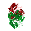
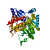
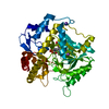
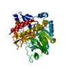
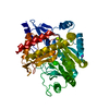
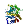
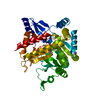
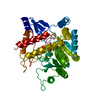
| Title | The crystal structure of dialkylglycine decarboxylase complex with pyridoxamine 5-phosphate | ||||||
 Components Components | 2,2-dialkylglycine decarboxylase | ||||||
 Keywords Keywords | LYASE / DGD complex with Pyridoxal 5-phosphate | ||||||
| Function / homology |  Function and homology information Function and homology information2,2-dialkylglycine decarboxylase (pyruvate) / 2,2-dialkylglycine decarboxylase (pyruvate) activity / transaminase activity / pyridoxal phosphate binding Similarity search - Function | ||||||
| Biological species |  Burkholderia cepacia (bacteria) Burkholderia cepacia (bacteria) | ||||||
| Method |  X-RAY DIFFRACTION / MOLECULAR REPLACEMENT / Resolution: 2 Å X-RAY DIFFRACTION / MOLECULAR REPLACEMENT / Resolution: 2 Å | ||||||
 Authors Authors | Fogle, E.J. / Liu, W. / Toney, M.D. | ||||||
 Citation Citation | Journal: Biochemistry / Year: 2005 Title: Role of q52 in catalysis of decarboxylation and transamination in dialkylglycine decarboxylase. Authors: Fogle, E.J. / Liu, W. / Woon, S.T. / Keller, J.W. / Toney, M.D. | ||||||
| History |
| ||||||
| Remark 999 | SEQUENCE As indicated by authors,the first residue methionine is missing in the database and the ... SEQUENCE As indicated by authors,the first residue methionine is missing in the database and the two other residues that conflict with the database are correct. The discrepancy is due to a mistake in the database when the protein was first sequenced. |
- Structure visualization
| Structure viewer | Molecule: MolmilJmol/JSmol |
|---|
- Downloads & links
-Download
| PDBx/mmCIF format | 1zc9.cif.gz | 102.5 KB | Display | PDBx/mmCIF format |
|---|---|---|---|---|
| PDB format | pdb1zc9.ent.gz | 76.7 KB | Display | PDB format |
| PDBx/mmJSON format | 1zc9.json.gz | Tree view | PDBx/mmJSON format | |
| Others |  Other downloads Other downloads |
-Validation report
| Arichive directory | https://data.pdbj.org/pub/pdb/validation_reports/zc/1zc9ftp://data.pdbj.org/pub/pdb/validation_reports/zc/1zc9 | HTTPS FTP |
|---|
-Related structure data
| Related structure data |  1z3zC 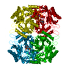 1dkaS S: Starting model for refinement C: citing same article ( |
|---|---|
| Similar structure data |


-Links
 PDBj
PDBj- Assembly
| Deposited unit | 
| ||||||||
|---|---|---|---|---|---|---|---|---|---|
| 1 |
| ||||||||
| Unit cell |
|
-Components
| #1: Protein | Mass: 46577.402 Da / Num. of mol.: 1 Source method: isolated from a genetically manipulated source Source: (gene. exp.) Burkholderia cepacia (bacteria) / Plasmid: JM109 / Production host: References: UniProt: P16932, 2,2-dialkylglycine decarboxylase (pyruvate) |
|---|---|
| #2: Chemical | ChemComp-K /   Mass: 39.098 Da / Num. of mol.: 1 / Source method: obtained synthetically / Formula: K Mass: 39.098 Da / Num. of mol.: 1 / Source method: obtained synthetically / Formula: K |
| #3: Chemical | ChemComp-NA /   Mass: 22.990 Da / Num. of mol.: 1 / Source method: obtained synthetically / Formula: Na Mass: 22.990 Da / Num. of mol.: 1 / Source method: obtained synthetically / Formula: Na |
| #4: Chemical | ChemComp-PMP /   Mass: 248.173 Da / Num. of mol.: 1 / Source method: obtained synthetically / Formula: C8H13N2O5P Mass: 248.173 Da / Num. of mol.: 1 / Source method: obtained synthetically / Formula: C8H13N2O5P |
| #5: Water | ChemComp-HOH /  Mass: 18.015 Da / Num. of mol.: 291 / Source method: isolated from a natural source / Formula: H2O Mass: 18.015 Da / Num. of mol.: 291 / Source method: isolated from a natural source / Formula: H2O |
-Experimental details
-Experiment
| Experiment | Method: X-RAY DIFFRACTION / Number of used crystals: 1 |
|---|
- Sample preparation
| Crystal | Density Matthews: 3.03 Å3/Da / Density % sol: 65 % |
|---|---|
| Crystal grow | Temperature: 298 K / Method: vapor diffusion, hanging drop / pH: 6.4 Details: MES, PEG4000, PLP, pH 6.4, VAPOR DIFFUSION, HANGING DROP, temperature 298K |
-Data collection
| Diffraction | Mean temperature: 100 K |
|---|---|
| Diffraction source | Source: ROTATING ANODE / Type: BRUKER / Wavelength: 1.54 Å |
| Detector | Type: BRUKER / Detector: CCD / Date: Jun 4, 2004 |
| Radiation | Monochromator: Ni / Protocol: SINGLE WAVELENGTH / Monochromatic (M) / Laue (L): M / Scattering type: x-ray |
| Radiation wavelength | Wavelength: 1.54 Å / Relative weight: 1 |
| Reflection | Resolution: 2→50 Å / Num. all: 39335 / Num. obs: 38428 / % possible obs: 97.7 % / Observed criterion σ(F): 2 / Observed criterion σ(I): 2 |
| Reflection shell | Resolution: 2→2.09 Å / % possible all: 97.4 |
- Processing
| Software |
| |||||||||||||||||||||||||
|---|---|---|---|---|---|---|---|---|---|---|---|---|---|---|---|---|---|---|---|---|---|---|---|---|---|---|
| Refinement | Method to determine structure: MOLECULAR REPLACEMENT Starting model: 1DKA Resolution: 2→50 Å / σ(F): 2 / Stereochemistry target values: Engh & Huber
| |||||||||||||||||||||||||
| Refinement step | Cycle: LAST / Resolution: 2→50 Å
| |||||||||||||||||||||||||
| Refine LS restraints |
| |||||||||||||||||||||||||
| Xplor file |
|
