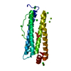
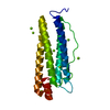
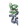
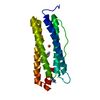
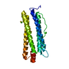
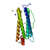
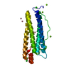
Entry Database : PDB / ID : 1yyhTitle Crystal structure of the human Notch 1 ankyrin domain Notch 1, ankyrin domain Keywords / / / Function / homology Function Domain/homology Component
/ / / / / / / / / / / / / / / / / / / / / / / / / / / / / / / / / / / / / / / / / / / / / / / / / / / / / / / / / / / / / / / / / / / / / / / / / / / / / / / / / / / / / / / / / / / / / / / / / / / / / / / / / / / / / / / / / / / / / / / / / / / / / / / / / / / / / / / / / / / / / / / / / / / / / / / / / / / / / / / / / Biological species Homo sapiens (human)Method / / / Resolution : 1.901 Å Authors Ehebauer, M.T. / Chirgadze, D.Y. / Hayward, P. / Martinez-Arias, A. / Blundell, T.L. Journal : Biochem.J. / Year : 2005Title : High-resolution crystal structure of the human Notch 1 ankyrin domainAuthors : Ehebauer, M.T. / Chirgadze, D.Y. / Hayward, P. / Martinez-Arias, A. / Blundell, T.L. History Deposition Feb 25, 2005 Deposition site / Processing site Revision 1.0 Aug 16, 2005 Provider / Type Revision 1.1 Apr 30, 2008 Group Revision 1.2 Jul 13, 2011 Group Revision 1.3 Jul 18, 2018 Group / Database references / Category Revision 1.4 Aug 23, 2023 Group / Database references / Refinement descriptionCategory chem_comp_atom / chem_comp_bond ... chem_comp_atom / chem_comp_bond / database_2 / pdbx_initial_refinement_model / struct_ref_seq_dif Item / _database_2.pdbx_database_accession / _struct_ref_seq_dif.details
Show all Show less
 Movie
Movie Controller


 Open data
 Basic information
 Components
 Keywords
 Function and homology information
 Homo sapiens (human)
 X-RAY DIFFRACTION /
  Authors
 Citation
 Structure visualization
 Downloads & links
 Other downloads

 PDBj
PDBj


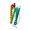
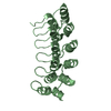
 Assembly

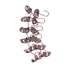
 Mass: 18.015 Da / Num. of mol.: 537 / Source method: isolated from a natural source / Formula: H2O
 Sample preparation
 / Beamline: PX14.1 / Wavelength: 1.488 Å
 Processing