 Movie
Movie Controller

[English] 日本語
 Yorodumi
- PDB-1yhu: Crystal structure of Riftia pachyptila C1 hemoglobin reveals nove... -
+ Open data

- Basic information
| Entry | Database: PDB / ID: 1yhu | ||||||
|---|---|---|---|---|---|---|---|
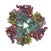
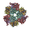
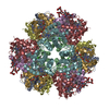
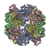
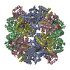
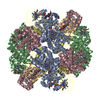
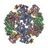
| Title | Crystal structure of Riftia pachyptila C1 hemoglobin reveals novel assembly of 24 subunits. | ||||||
 Components Components |
| ||||||
 Keywords Keywords | OXYGEN STORAGE/TRANSPORT / hemoglobin / globin fold / OXYGEN STORAGE-TRANSPORT COMPLEX | ||||||
| Function / homology |  Function and homology information Function and homology informationhemoglobin complex / oxygen carrier activity / oxygen binding / iron ion binding / heme binding / extracellular space / extracellular region Similarity search - Function | ||||||
| Biological species |  Riftia pachyptila (tube worm) Riftia pachyptila (tube worm) | ||||||
| Method |  X-RAY DIFFRACTION / SYNCHROTRON / Plus molecular averaging / Resolution: 3.15 Å X-RAY DIFFRACTION / SYNCHROTRON / Plus molecular averaging / Resolution: 3.15 Å | ||||||
 Authors Authors | Flores, J.F. / Fisher, C.R. / Carney, S.L. / Green, B.N. / Freytag, J.K. / Schaeffer, S.W. / Royer, W.E. | ||||||
 Citation Citation | Journal: Proc.Natl.Acad.Sci.Usa / Year: 2005 Title: Sulfide binding is mediated by zinc ions discovered in the crystal structure of a hydrothermal vent tubeworm hemoglobin. Authors: Flores, J.F. / Fisher, C.R. / Carney, S.L. / Green, B.N. / Freytag, J.K. / Schaeffer, S.W. / Royer Jr, W.E. | ||||||
| History |
|
- Structure visualization
| Structure viewer | Molecule: MolmilJmol/JSmol |
|---|
- Downloads & links
-Download
| PDBx/mmCIF format | 1yhu.cif.gz | 624.6 KB | Display | PDBx/mmCIF format |
|---|---|---|---|---|
| PDB format | pdb1yhu.ent.gz | 543.5 KB | Display | PDB format |
| PDBx/mmJSON format | 1yhu.json.gz | Tree view | PDBx/mmJSON format | |
| Others |  Other downloads Other downloads |
-Validation report
| Summary document | 1yhu_validation.pdf.gz | 6.7 MB | Display | wwPDB validaton report |
|---|---|---|---|---|
| Full document | 1yhu_full_validation.pdf.gz | 6.9 MB | Display | |
| Data in XML | 1yhu_validation.xml.gz | 141.2 KB | Display | |
| Data in CIF | 1yhu_validation.cif.gz | 176.3 KB | Display | |
| Arichive directory | https://data.pdbj.org/pub/pdb/validation_reports/yh/1yhuftp://data.pdbj.org/pub/pdb/validation_reports/yh/1yhu | HTTPS FTP |
-Related structure data
| Similar structure data |
|---|

-Links
 PDBj
PDBj


- Assembly
| Deposited unit | 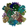
| ||||||||
|---|---|---|---|---|---|---|---|---|---|
| 1 |
| ||||||||
| Unit cell |
| ||||||||
| Details | 24mer assembled with D3 symmetry relating A1A2B1B2 tetramer. One entire assembly is present in the asymmetric unit. |
-Components
-Protein , 4 types, 24 molecules AEIMQUBFJNRVCGKOSWDHLPTX
| #1: Protein | Mass: 15795.024 Da / Num. of mol.: 6 / Source method: isolated from a natural source / Source: (natural) Riftia pachyptila (tube worm) / References: UniProt: Q8IFK4#2: Protein | Mass: 16158.232 Da / Num. of mol.: 6 / Source method: isolated from a natural source / Source: (natural) Riftia pachyptila (tube worm) / References: UniProt: P80592#3: Protein | Mass: 15285.326 Da / Num. of mol.: 6 / Source method: isolated from a natural source / Source: (natural) Riftia pachyptila (tube worm) / References: UniProt: Q8IFK2#4: Protein | Mass: 16162.318 Da / Num. of mol.: 6 / Source method: isolated from a natural source / Source: (natural) Riftia pachyptila (tube worm) / References: UniProt: Q8IFJ9 |
|---|
-Non-polymers , 3 types, 60 molecules 


| #5: Chemical | ChemComp-HEM /  Mass: 616.487 Da / Num. of mol.: 24 / Source method: obtained synthetically / Formula: C34H32FeN4O4 Mass: 616.487 Da / Num. of mol.: 24 / Source method: obtained synthetically / Formula: C34H32FeN4O4#6: Chemical | ChemComp-OXY /  Mass: 31.999 Da / Num. of mol.: 24 / Source method: obtained synthetically / Formula: O2 Mass: 31.999 Da / Num. of mol.: 24 / Source method: obtained synthetically / Formula: O2#7: Chemical | ChemComp-ZN /  Mass: 65.409 Da / Num. of mol.: 12 / Source method: obtained synthetically / Formula: Zn Mass: 65.409 Da / Num. of mol.: 12 / Source method: obtained synthetically / Formula: Zn |
|---|
-Experimental details
-Experiment
| Experiment | Method: X-RAY DIFFRACTION / Number of used crystals: 1 |
|---|
- Sample preparation
| Crystal | Density Matthews: 3.89 Å3/Da / Density % sol: 68.4 % |
|---|---|
| Crystal grow | Temperature: 293 K / Method: vapor diffusion, sitting drop / pH: 7.5 Details: 1.6M ammonium sulfate, 0.1M HEPES, 0.1M NaCl, pH 7.5, VAPOR DIFFUSION, SITTING DROP, temperature 293K |
-Data collection
| Diffraction | Mean temperature: 100 K |
|---|---|
| Diffraction source | Source: SYNCHROTRON / Site: APS  / Beamline: 14-BM-C / Wavelength: 0.9 Å / Beamline: 14-BM-C / Wavelength: 0.9 Å |
| Detector | Type: ADSC QUANTUM 4 / Detector: CCD / Date: Dec 10, 2002 |
| Radiation | Protocol: SINGLE WAVELENGTH / Monochromatic (M) / Laue (L): M / Scattering type: x-ray |
| Radiation wavelength | Wavelength: 0.9 Å / Relative weight: 1 |
| Reflection | Resolution: 3.15→50 Å / Num. all: 104444 / Num. obs: 104444 / % possible obs: 100 % / Observed criterion σ(F): 0 / Observed criterion σ(I): 0 / Redundancy: 4.6 % / Rsym value: 0.065 / Net I/σ(I): 11.3 |
| Reflection shell | Resolution: 3.15→3.26 Å / Mean I/σ(I) obs: 2.4 / Rsym value: 0.316 / % possible all: 99.9 |
- Processing
| Software |
| ||||||||||||||||||||
|---|---|---|---|---|---|---|---|---|---|---|---|---|---|---|---|---|---|---|---|---|---|
| Refinement | Method to determine structure: Plus molecular averaging / Resolution: 3.15→50 Å / σ(F): 0 / Stereochemistry target values: Engh & Huber
| ||||||||||||||||||||
| Refinement step | Cycle: LAST / Resolution: 3.15→50 Å
|
