 Movie
Movie Controller

[English] 日本語
 Yorodumi
- PDB-1yfi: Crystal Structure of restriction endonuclease MspI in complex wit... -
+ Open data

- Basic information
| Entry | Database: PDB / ID: 1yfi | ||||||
|---|---|---|---|---|---|---|---|
| Title | Crystal Structure of restriction endonuclease MspI in complex with its cognate DNA in P212121 space group | ||||||
 Components Components |
| ||||||
 Keywords Keywords | Hydrolase/DNA / protein-DNA complex / Hydrolase-DNA COMPLEX | ||||||
| Function / homology | Restriction endonuclease, type II, MspI / Restriction endonuclease MspI / type II site-specific deoxyribonuclease / type II site-specific deoxyribonuclease activity / Restriction endonuclease type II-like / DNA restriction-modification system / DNA binding / DNA / Type II restriction enzyme MspI Function and homology information Function and homology information | ||||||
| Biological species |  Moraxella sp. (bacteria) Moraxella sp. (bacteria) | ||||||
| Method |  X-RAY DIFFRACTION / SYNCHROTRON / MOLECULAR REPLACEMENT / Resolution: 2.7 Å X-RAY DIFFRACTION / SYNCHROTRON / MOLECULAR REPLACEMENT / Resolution: 2.7 Å | ||||||
 Authors Authors | Xu, Q.S. / Kucera, R.B. / Roberts, R.J. / Guo, H.-C. | ||||||
 Citation Citation | Journal: Protein Sci. / Year: 2005 Title: Two crystal forms of the restriction enzyme MspI-DNA complex show the same novel structure. Authors: Xu, Q.S. / Roberts, R.J. / Guo, H.-C. | ||||||
| History |
|
- Structure visualization
| Structure viewer | Molecule: MolmilJmol/JSmol |
|---|
- Downloads & links
-Download
| PDBx/mmCIF format | 1yfi.cif.gz | 136.5 KB | Display | PDBx/mmCIF format |
|---|---|---|---|---|
| PDB format | pdb1yfi.ent.gz | 104.5 KB | Display | PDB format |
| PDBx/mmJSON format | 1yfi.json.gz | Tree view | PDBx/mmJSON format | |
| Others |  Other downloads Other downloads |
-Validation report
| Arichive directory | https://data.pdbj.org/pub/pdb/validation_reports/yf/1yfiftp://data.pdbj.org/pub/pdb/validation_reports/yf/1yfi | HTTPS FTP |
|---|
-Related structure data
| Related structure data |  1sa3S S: Starting model for refinement |
|---|---|
| Similar structure data |

-Links
 PDBj
PDBj


- Assembly
| Deposited unit | 
| ||||||||
|---|---|---|---|---|---|---|---|---|---|
| 1 | 
| ||||||||
| 2 | 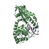
| ||||||||
| Unit cell |
|
-Components
| #1: DNA chain | Mass: 3046.980 Da / Num. of mol.: 4 / Source method: obtained synthetically #2: Protein | Mass: 29876.010 Da / Num. of mol.: 2 Source method: isolated from a genetically manipulated source Source: (gene. exp.) Moraxella sp. (bacteria) / Gene: mspIR / Plasmid: PCAD39 / Production host: References: UniProt: P11405, type II site-specific deoxyribonuclease #3: Water | ChemComp-HOH / |  Mass: 18.015 Da / Num. of mol.: 53 / Source method: isolated from a natural source / Formula: H2O Mass: 18.015 Da / Num. of mol.: 53 / Source method: isolated from a natural source / Formula: H2O |
|---|
-Experimental details
-Experiment
| Experiment | Method: X-RAY DIFFRACTION / Number of used crystals: 1 |
|---|
- Sample preparation
| Crystal | Density Matthews: 2.58 Å3/Da / Density % sol: 52 % | ||||||||||||||||||||||||||||||||||||||||
|---|---|---|---|---|---|---|---|---|---|---|---|---|---|---|---|---|---|---|---|---|---|---|---|---|---|---|---|---|---|---|---|---|---|---|---|---|---|---|---|---|---|
| Crystal grow | Temperature: 298 K / Method: vapor diffusion, hanging drop / pH: 6.5 Details: MES, ammonium sulfate, calcium chloride, PEG 8000, glycerol , pH 6.5, VAPOR DIFFUSION, HANGING DROP, temperature 298K | ||||||||||||||||||||||||||||||||||||||||
| Components of the solutions |
|
-Data collection
| Diffraction | Mean temperature: 100 K |
|---|---|
| Diffraction source | Source: SYNCHROTRON / Site: NSLS  / Beamline: X12C / Wavelength: 0.904 Å / Beamline: X12C / Wavelength: 0.904 Å |
| Detector | Type: BRANDEIS - B4 / Detector: CCD / Date: Apr 8, 2001 Details: DOUBLE-CRYSTAL MONOCHROMATOR SI(111), BEAM FOCUSED BY A TOROIDAL MIRROR |
| Radiation | Monochromator: DOUBLE-CRYSTAL MONOCHROMATOR Si(111) MIRROR + NI FILTER Protocol: SINGLE WAVELENGTH / Monochromatic (M) / Laue (L): M / Scattering type: x-ray |
| Radiation wavelength | Wavelength: 0.904 Å / Relative weight: 1 |
| Reflection | Resolution: 2.7→100 Å / Num. all: 21426 / Num. obs: 21426 / % possible obs: 99.9 % / Observed criterion σ(F): 0 / Observed criterion σ(I): -3 / Redundancy: 10 % / Biso Wilson estimate: 27.9 Å2 / Rmerge(I) obs: 0.072 / Rsym value: 0.072 / Net I/σ(I): 28.7 |
| Reflection shell | Resolution: 2.7→2.8 Å / Rmerge(I) obs: 0.282 / Mean I/σ(I) obs: 7 / Num. unique all: 2100 / Rsym value: 0.282 / % possible all: 100 |
- Processing
| Software |
| ||||||||||||||||||||||||||||||||||||
|---|---|---|---|---|---|---|---|---|---|---|---|---|---|---|---|---|---|---|---|---|---|---|---|---|---|---|---|---|---|---|---|---|---|---|---|---|---|
| Refinement | Method to determine structure: MOLECULAR REPLACEMENT Starting model: PDB ENTRY 1SA3 Resolution: 2.7→46.94 Å / Rfactor Rfree error: 0.006 / Data cutoff high absF: 1435032.38 / Data cutoff high rms absF: 1435032.38 / Data cutoff low absF: 0 / Isotropic thermal model: RESTRAINED / Cross valid method: THROUGHOUT / σ(F): 0 / Stereochemistry target values: Engh & Huber
| ||||||||||||||||||||||||||||||||||||
| Solvent computation | Solvent model: FLAT MODEL / Bsol: 10 Å2 / ksol: 0.318155 e/Å3 | ||||||||||||||||||||||||||||||||||||
| Displacement parameters | Biso mean: 26.7 Å2
| ||||||||||||||||||||||||||||||||||||
| Refine analyze |
| ||||||||||||||||||||||||||||||||||||
| Refinement step | Cycle: LAST / Resolution: 2.7→46.94 Å
| ||||||||||||||||||||||||||||||||||||
| Refine LS restraints |
| ||||||||||||||||||||||||||||||||||||
| LS refinement shell | Resolution: 2.7→2.8 Å / Rfactor Rfree error: 0.027 / Total num. of bins used: 10
| ||||||||||||||||||||||||||||||||||||
| Xplor file |
|
