 Movie
Movie Controller

[English] 日本語
 Yorodumi
- PDB-1xpn: NMR structure of P. aeruginosa protein PA1324: Northeast Structur... -
+ Open data

- Basic information
| Entry | Database: PDB / ID: 1xpn | ||||||
|---|---|---|---|---|---|---|---|
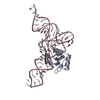
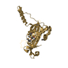
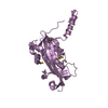
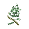
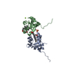
| Title | NMR structure of P. aeruginosa protein PA1324: Northeast Structural Genomics Consortium target PaP1 | ||||||
 Components Components | hypothetical protein PA1324 | ||||||
 Keywords Keywords | STRUCTURAL GENOMICS / UNKNOWN FUNCTION / b-barrel / Northeast Structural Genomics Consortium / NESG / Protein Structure Initiative / PSI | ||||||
| Function / homology | Prokaryotic membrane lipoprotein lipid attachment site profile. / Carboxypeptidase regulatory-like domain-containing protein Function and homology information Function and homology information | ||||||
| Biological species |   Pseudomonas aeruginosa (bacteria) Pseudomonas aeruginosa (bacteria) | ||||||
| Method | SOLUTION NMR / Molecular Dynamics, Simulated Annealing | ||||||
 Authors Authors | Cort, J.R. / Ni, S. / Lockert, E.E. / Montelione, G.T. / Kennedy, M.A. / Northeast Structural Genomics Consortium (NESG) | ||||||
 Citation Citation | Journal: Protein Sci. / Year: 2009 Title: Structure and function of Pseudomonas aeruginosa protein PA1324 (21-170). Authors: Mercier, K.A. / Cort, J.R. / Kennedy, M.A. / Lockert, E.E. / Ni, S. / Shortridge, M.D. / Powers, R. | ||||||
| History |
|
- Structure visualization
| Structure viewer | Molecule: MolmilJmol/JSmol |
|---|
- Downloads & links
-Download
| PDBx/mmCIF format | 1xpn.cif.gz | 986.6 KB | Display | PDBx/mmCIF format |
|---|---|---|---|---|
| PDB format | pdb1xpn.ent.gz | 826.3 KB | Display | PDB format |
| PDBx/mmJSON format | 1xpn.json.gz | Tree view | PDBx/mmJSON format | |
| Others |  Other downloads Other downloads |
-Validation report
| Arichive directory | https://data.pdbj.org/pub/pdb/validation_reports/xp/1xpnftp://data.pdbj.org/pub/pdb/validation_reports/xp/1xpn | HTTPS FTP |
|---|
-Related structure data
| Similar structure data | |
|---|---|
| Other databases |


-Links
 PDBj
PDBj


- Assembly
| Deposited unit | 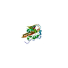
| |||||||||
|---|---|---|---|---|---|---|---|---|---|---|
| 1 |
| |||||||||
| NMR ensembles |
|
-Components
| #1: Protein | Mass: 18403.201 Da / Num. of mol.: 1 Source method: isolated from a genetically manipulated source Source: (gene. exp.) Pseudomonas aeruginosa (bacteria) / Strain: PAO1 / Gene: PA1324 / Species (production host): Escherichia coli / Production host: |
|---|
-Experimental details
-Experiment
| Experiment | Method: SOLUTION NMR | ||||||||||||||||
|---|---|---|---|---|---|---|---|---|---|---|---|---|---|---|---|---|---|
| NMR experiment |
|
- Sample preparation
| Details |
| |||||||||
|---|---|---|---|---|---|---|---|---|---|---|
| Sample conditions | Ionic strength: 300 mM NaCl / pH: 6.8 / Pressure: ambient / Temperature: 298 K |
-NMR measurement
| Radiation | Protocol: SINGLE WAVELENGTH / Monochromatic (M) / Laue (L): M / Scattering type: x-ray | ||||||||||||||||||||
|---|---|---|---|---|---|---|---|---|---|---|---|---|---|---|---|---|---|---|---|---|---|
| Radiation wavelength | Relative weight: 1 | ||||||||||||||||||||
| NMR spectrometer |
|
- Processing
| NMR software |
| ||||||||||||||||||||||||
|---|---|---|---|---|---|---|---|---|---|---|---|---|---|---|---|---|---|---|---|---|---|---|---|---|---|
| Refinement | Method: Molecular Dynamics, Simulated Annealing / Software ordinal: 1 Details: NOE distance restraints were determined automatically using AutoStructure (G.T. Montelione & Y.J. Huang) starting from peak-picked NOESY data and chemical shift assignments. Final refinement ...Details: NOE distance restraints were determined automatically using AutoStructure (G.T. Montelione & Y.J. Huang) starting from peak-picked NOESY data and chemical shift assignments. Final refinement was conducted in explicit solvent with Leonard-Jones and electrostatic potentials. Residues 1-26 are unrestrained and constitute a flexible, n-terminal tail. The structure of residues 27-170 is restrained by 1861 noe distance restraints (541 intraresidue, 513 sequential, 190 medium range, and 617 long range (n>4)), 132 dihedral restraints (65 phi, 67 psi), and 70 h-bond restraints (for 35 h-bonds). Dihedral restraints were derived from Talos and HNHA. H-bond restraints were derived from analysis of amides that exchange slowly with D2O solvent together with initial structures derived from Noesy data. | ||||||||||||||||||||||||
| NMR representative | Selection criteria: lowest energy, fewest violations, closest to the average | ||||||||||||||||||||||||
| NMR ensemble | Conformer selection criteria: structures with the lowest energy,structures with the fewest restraint violations Conformers calculated total number: 20 / Conformers submitted total number: 20 |
