 Movie
Movie Controller

[English] 日本語
 Yorodumi
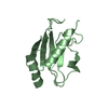
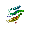
- PDB-1xg8: Crystal Structure of Protein of Unknown Function SA0789 from Stap... -
+ Open data

- Basic information
| Entry | Database: PDB / ID: 1xg8 | ||||||
|---|---|---|---|---|---|---|---|
| Title | Crystal Structure of Protein of Unknown Function SA0789 from Staphylococcus aureus | ||||||
 Components Components | hypothetical protein SA0798 | ||||||
 Keywords Keywords | STRUCTURAL GENOMICS / UNKNOWN FUNCTION / Protein Structure Initative / MCSG / Staphylococcus aureus / PSI / Protein Structure Initiative / Midwest Center for Structural Genomics | ||||||
| Function / homology |  Function and homology information Function and homology informationHypothetical protein sa0798. / Protein of unknown function DUF1462 / YuzD-like superfamily / Protein of unknown function (DUF1462) / Glutaredoxin / Thioredoxin-like superfamily / 3-Layer(aba) Sandwich / Alpha Beta Similarity search - Domain/homology | ||||||
| Biological species |  Staphylococcus aureus subsp. aureus N315 (bacteria) Staphylococcus aureus subsp. aureus N315 (bacteria) | ||||||
| Method |  X-RAY DIFFRACTION / SYNCHROTRON / MAD / Resolution: 2.1 Å X-RAY DIFFRACTION / SYNCHROTRON / MAD / Resolution: 2.1 Å | ||||||
 Authors Authors | Rotella, F.J. / Zhang, R.G. / Kim, Y. / Quartey, P. / Collart, F. / Joachimiak, A. / Midwest Center for Structural Genomics (MCSG) | ||||||
 Citation Citation | Journal: To be Published Title: The 2.1A crystal structure of hypothetical protein SA0798 from Staphylococcus aureus Authors: Rotella, F.J. / Zhang, R.G. / Kim, Y. / Quartey, P. / Collart, F. / Joachimiak, A. | ||||||
| History |
|
- Structure visualization
| Structure viewer | Molecule: MolmilJmol/JSmol |
|---|
- Downloads & links
-Download
| PDBx/mmCIF format | 1xg8.cif.gz | 34.8 KB | Display | PDBx/mmCIF format |
|---|---|---|---|---|
| PDB format | pdb1xg8.ent.gz | 23.4 KB | Display | PDB format |
| PDBx/mmJSON format | 1xg8.json.gz | Tree view | PDBx/mmJSON format | |
| Others |  Other downloads Other downloads |
-Validation report
| Summary document | 1xg8_validation.pdf.gz | 428 KB | Display | wwPDB validaton report |
|---|---|---|---|---|
| Full document | 1xg8_full_validation.pdf.gz | 431.2 KB | Display | |
| Data in XML | 1xg8_validation.xml.gz | 7.1 KB | Display | |
| Data in CIF | 1xg8_validation.cif.gz | 8.8 KB | Display | |
| Arichive directory | https://data.pdbj.org/pub/pdb/validation_reports/xg/1xg8ftp://data.pdbj.org/pub/pdb/validation_reports/xg/1xg8 | HTTPS FTP |
-Related structure data
| Similar structure data | |
|---|---|
| Other databases |


-Links
 PDBj
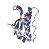
PDBj- Assembly
| Deposited unit | 
| ||||||||
|---|---|---|---|---|---|---|---|---|---|
| 1 |
| ||||||||
| Unit cell |
| ||||||||
| Details | the biological assembly is monomer |
-Components
| #1: Protein | Mass: 13038.609 Da / Num. of mol.: 1 Source method: isolated from a genetically manipulated source Source: (gene. exp.) Staphylococcus aureus subsp. aureus N315 (bacteria)Species: Staphylococcus aureus / Strain: subsp. aureus N315 / Gene: GI:1123613 / Plasmid: PDM68 / Species (production host): Escherichia coli / Production host: |
|---|---|
| #2: Water | ChemComp-HOH /  Mass: 18.015 Da / Num. of mol.: 41 / Source method: isolated from a natural source / Formula: H2O Mass: 18.015 Da / Num. of mol.: 41 / Source method: isolated from a natural source / Formula: H2O |
-Experimental details
-Experiment
| Experiment | Method: X-RAY DIFFRACTION / Number of used crystals: 1 |
|---|
- Sample preparation
| Crystal | Density Matthews: 2.35 Å3/Da / Density % sol: 45.8 % |
|---|---|
| Crystal grow | Temperature: 273 K / Method: vapor diffusion, sitting drop / pH: 5 Details: ammonium sulfate, citrate buffer, pH 5, VAPOR DIFFUSION, SITTING DROP, temperature 273K |
-Data collection
| Diffraction | Mean temperature: 100 K | ||||||||||||
|---|---|---|---|---|---|---|---|---|---|---|---|---|---|
| Diffraction source | Source: SYNCHROTRON / Site: APS  / Beamline: 19-ID / Wavelength: 0.979569, 0.979706, 0.964107 / Beamline: 19-ID / Wavelength: 0.979569, 0.979706, 0.964107 | ||||||||||||
| Detector | Type: SBC-2 / Detector: CCD / Date: Mar 16, 2004 / Details: vertical-focusing mirror | ||||||||||||
| Radiation | Monochromator: Si-111 double-crystal channel / Protocol: MAD / Monochromatic (M) / Laue (L): M / Scattering type: x-ray | ||||||||||||
| Radiation wavelength |
| ||||||||||||
| Reflection | Resolution: 2.1→50 Å / Num. all: 12274 / Num. obs: 11783 / % possible obs: 96 % / Observed criterion σ(F): 2 / Observed criterion σ(I): 2 / Redundancy: 7 % / Biso Wilson estimate: 14.5 Å2 / Rmerge(I) obs: 0.083 / Net I/σ(I): 18.8 | ||||||||||||
| Reflection shell | Resolution: 2.1→2.18 Å / Redundancy: 2.7 % / Rmerge(I) obs: 0.481 / Mean I/σ(I) obs: 1.73 / Num. unique all: 1235 / % possible all: 71.8 |
- Processing
| Software |
| |||||||||||||||||||||||||
|---|---|---|---|---|---|---|---|---|---|---|---|---|---|---|---|---|---|---|---|---|---|---|---|---|---|---|
| Refinement | Method to determine structure: MAD / Resolution: 2.1→29.6 Å / Rfactor Rfree error: 0.012 / Data cutoff high absF: 226196.73 / Data cutoff low absF: 0 / Isotropic thermal model: RESTRAINED / Cross valid method: THROUGHOUT / σ(F): 0 / Stereochemistry target values: Engh & Huber
| |||||||||||||||||||||||||
| Solvent computation | Solvent model: FLAT MODEL / Bsol: 60.2014 Å2 / ksol: 0.39592 e/Å3 | |||||||||||||||||||||||||
| Displacement parameters | Biso mean: 30.5 Å2
| |||||||||||||||||||||||||
| Refine analyze |
| |||||||||||||||||||||||||
| Refinement step | Cycle: LAST / Resolution: 2.1→29.6 Å
| |||||||||||||||||||||||||
| Refine LS restraints |
| |||||||||||||||||||||||||
| LS refinement shell | Resolution: 2.1→2.23 Å / Rfactor Rfree error: 0.045 / Total num. of bins used: 6
| |||||||||||||||||||||||||
| Xplor file |
|
