- PDB-1xdq: Structural and Biochemical Identification of a Novel Bacterial Ox... -
+
Open data
ID or keywords:
Loading...
-
Basic information
Entry
Database: PDB / ID: 1xdq
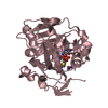
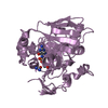
Title
Structural and Biochemical Identification of a Novel Bacterial Oxidoreductase
Components
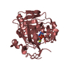
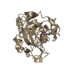
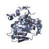
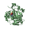
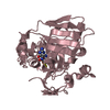
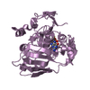
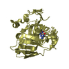
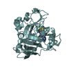
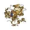
Bacterial Sulfite Oxidase
Keywords
OXIDOREDUCTASE / Bioinformatics / sequence analysis / electron transfer / molybdoenzymes / molybdopterin
Function / homology
Function and homology information
Oxidoreductases; Acting on a sulfur group of donors; With a quinone or similar compound as acceptor / oxidoreductase activity, acting on a heme group of donors / response to hypochlorite / oxidoreductase activity, acting on a sulfur group of donors, quinone or similar compound as acceptor / protein repair / molybdopterin cofactor binding / outer membrane-bounded periplasmic space / oxidoreductase activity / metal ion binding Similarity search - Function
HETEROGEN Due to the presence of adjacent electron density from a bound molecule, we cannot ...HETEROGEN Due to the presence of adjacent electron density from a bound molecule, we cannot distinguish unambiguously the position of the second coordinating atom as it can be refined with similar parameters at the typical length for an oxo group (1.6-1.8 and a direct hydrogen bond to bound molecule) or for a water molecule (2.1-2.4 and a direct hydrogen bond to bound urea).
Remark 999
SEQUENCE THE AUTHORS BELIEVE THAT THERE IS AN AN ERROR IN THE DATABASE SEQUENCE SINCE THE OTHER E. ...SEQUENCE THE AUTHORS BELIEVE THAT THERE IS AN AN ERROR IN THE DATABASE SEQUENCE SINCE THE OTHER E.COLI STRAINS WITH PUBLISHED SEQUENCES LIKE THE PATHOGENIC STRAINS O157:H7 EDL933, AND CFT073 ALSO HAVE AN ASP AT THAT POSITION.
In the structure databanks used in Yorodumi, some data are registered as the other names, "COVID-19 virus" and "2019-nCoV". Here are the details of the virus and the list of structure data.
Jan 31, 2019. EMDB accession codes are about to change! (news from PDBe EMDB page)
EMDB accession codes are about to change! (news from PDBe EMDB page)
The allocation of 4 digits for EMDB accession codes will soon come to an end. Whilst these codes will remain in use, new EMDB accession codes will include an additional digit and will expand incrementally as the available range of codes is exhausted. The current 4-digit format prefixed with “EMD-” (i.e. EMD-XXXX) will advance to a 5-digit format (i.e. EMD-XXXXX), and so on. It is currently estimated that the 4-digit codes will be depleted around Spring 2019, at which point the 5-digit format will come into force.
The EM Navigator/Yorodumi systems omit the EMD- prefix.
Related info.:Q: What is EMD? / ID/Accession-code notation in Yorodumi/EM Navigator
Yorodumi is a browser for structure data from EMDB, PDB, SASBDB, etc.
This page is also the successor to EM Navigator detail page, and also detail information page/front-end page for Omokage search.
The word "yorodu" (or yorozu) is an old Japanese word meaning "ten thousand". "mi" (miru) is to see.
Related info.:EMDB / PDB / SASBDB / Comparison of 3 databanks / Yorodumi Search / Aug 31, 2016. New EM Navigator & Yorodumi / Yorodumi Papers / Jmol/JSmol / Function and homology information / Changes in new EM Navigator and Yorodumi
 Movie
Movie Controller

 Yorodumi
 Open data
 Basic information
 Components
 Keywords
 Function and homology information
 X-RAY DIFFRACTION /
  Authors
 Citation
 Structure visualization
 Downloads & links
 Other downloads

 PDBj
PDBj


 Assembly


 Mass: 60.055 Da / Num. of mol.: 5 / Source method: obtained synthetically / Formula: CH4N2O
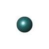
 Mass: 95.940 Da / Num. of mol.: 5 / Source method: obtained synthetically / Formula: Mo
 Mass: 15.999 Da / Num. of mol.: 5 / Source method: obtained synthetically / Formula: O
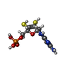
 Mass: 395.352 Da / Num. of mol.: 5 / Source method: obtained synthetically / Formula: C10H14N5O6PS2
 Sample preparation
 / Beamline: 8.3.1 / Wavelength: 0.97965 Å
 Processing