 Movie
Movie Controller


+ Open data

- Basic information
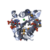
| Entry | Database: PDB / ID: 1xbs | ||||||
|---|---|---|---|---|---|---|---|
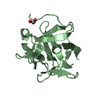
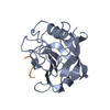
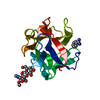
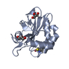
| Title | Crystal structure of human dim2: a dim1-like protein | ||||||
 Components Components | Dim1-like protein | ||||||
 Keywords Keywords |  TRANSCRIPTION / CELL CYCLE / SPLICEOSOMAL PROTEIN / THIOREDOXIN / SNRNP TRANSCRIPTION / CELL CYCLE / SPLICEOSOMAL PROTEIN / THIOREDOXIN / SNRNP | ||||||
| Function / homology |  Function and homology information Function and homology informationU5 snRNP / U4/U6 x U5 tri-snRNP complex / spliceosomal complex / mRNA splicing, via spliceosome / cell cycle / nucleoplasm / cytosolSimilarity search - Function | ||||||
| Biological species |  Homo sapiens (human) Homo sapiens (human) | ||||||
| Method | X-RAY DIFFRACTION / SYNCHROTRON / MOLECULAR REPLACEMENT / Resolution: 2.5 Å | ||||||
 Authors Authors | Simeoni, F. / Arvai, A. / Hopfner, K.-P. / Bello, P. / Gondeau, C. / Heitz, F. / Tainer, J. / Divita, G. | ||||||
 Citation Citation | Journal: Biochemistry / Year: 2005 Title: Biochemical Characterization and Crystal Structure of a Dim1 Family Associated Protein: Dim2 Authors: Simeoni, F. / Arvai, A. / Bello, P. / Gondeau, C. / Hopfner, K.-P. / Neyroz, P. / Heitz, F. / Tainer, J. / Divita, G. | ||||||
| History |
|
- Structure visualization
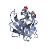
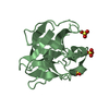
| Structure viewer | Molecule: MolmilJmol/JSmol |
|---|
- Downloads & links
-Download
| PDBx/mmCIF format | 1xbs.cif.gz | 38.3 KB | Display | PDBx/mmCIF format |
|---|---|---|---|---|
| PDB format | pdb1xbs.ent.gz | 26.9 KB | Display | PDB format |
| PDBx/mmJSON format | 1xbs.json.gz | Tree view | PDBx/mmJSON format | |
| Others |  Other downloads Other downloads |
-Validation report
| Arichive directory | https://data.pdbj.org/pub/pdb/validation_reports/xb/1xbsftp://data.pdbj.org/pub/pdb/validation_reports/xb/1xbs | HTTPS FTP |
|---|
-Related structure data
| Similar structure data |
|---|


-Links
 PDBj
PDBj- Assembly
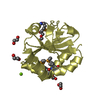
| Deposited unit | 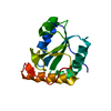
| ||||||||
|---|---|---|---|---|---|---|---|---|---|
| 1 |
| ||||||||
| Unit cell |
|
-Components
| #1: Protein | Mass: 17034.611 Da / Num. of mol.: 1 Source method: isolated from a genetically manipulated source Source: (gene. exp.) Homo sapiens (human) / Plasmid: pGEX4T / Production host:  Escherichia coli (E. coli) / Strain (production host): DH5a / References: GenBank: 45594298, UniProt: Q9NX01*PLUS Escherichia coli (E. coli) / Strain (production host): DH5a / References: GenBank: 45594298, UniProt: Q9NX01*PLUS |
|---|---|
| #2: Water | ChemComp-HOH / Water Mass: 18.015 Da / Num. of mol.: 21 / Source method: isolated from a natural source / Formula: H2O Mass: 18.015 Da / Num. of mol.: 21 / Source method: isolated from a natural source / Formula: H2O |
-Experimental details
-Experiment
| Experiment | Method: X-RAY DIFFRACTION / Number of used crystals: 1 |
|---|
- Sample preparation
| Crystal | Density Matthews: 2.98 Å3/Da / Density % sol: 58.4 % |
|---|---|
| Crystal grow | Temperature: 293 K / Method: vapor diffusion, hanging drop / pH: 7.2 Details: Ammonium sulfate, sodium chloride, EDTA, Tris-HCl, pH 7.2, VAPOR DIFFUSION, HANGING DROP, temperature 293K |
-Data collection
| Diffraction | Mean temperature: 200 K |
|---|---|
| Diffraction source | Source: SYNCHROTRON / Site: SSRL  / Beamline: BL7-1 / Wavelength: 1.08 Å / Beamline: BL7-1 / Wavelength: 1.08 Å |
| Detector | Type: MARRESEARCH / Detector: IMAGE PLATE / Date: Jun 23, 2000 |
| Radiation | Protocol: SINGLE WAVELENGTH / Monochromatic (M) / Laue (L): M / Scattering type: x-ray |
| Radiation wavelength | Wavelength: 1.08 Å / Relative weight: 1 |
| Reflection | Resolution: 2.5→30 Å / Num. all: 6836 / Num. obs: 6538 / % possible obs: 89.9 % / Observed criterion σ(F): 1 / Observed criterion σ(I): 1 |
| Reflection shell | Highest resolution: 2.5 Å / Num. unique all: 615 / % possible all: 93.8 |
- Processing
| Software |
| ||||||||||||||||||||
|---|---|---|---|---|---|---|---|---|---|---|---|---|---|---|---|---|---|---|---|---|---|
| Refinement | Method to determine structure: MOLECULAR REPLACEMENT / Resolution: 2.5→30 Å / σ(F): -3 / Stereochemistry target values: Engh & Huber
| ||||||||||||||||||||
| Refinement step | Cycle: LAST / Resolution: 2.5→30 Å
| ||||||||||||||||||||
| Refine LS restraints |
|
