 Movie
Movie Controller


+ Open data

- Basic information
| Entry | Database: PDB / ID: 1wuw | ||||||
|---|---|---|---|---|---|---|---|
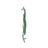
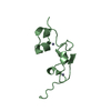
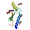
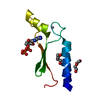
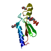
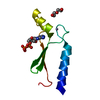
| Title | Crystal Structure of beta hordothionin | ||||||
 Components Components | Beta-hordothionin | ||||||
 Keywords Keywords | PLANT PROTEIN / crambin fold / dimer | ||||||
| Function / homology |  Function and homology information Function and homology information | ||||||
| Biological species |  | ||||||
| Method |  X-RAY DIFFRACTION / MOLECULAR REPLACEMENT / Resolution: 1.9 Å X-RAY DIFFRACTION / MOLECULAR REPLACEMENT / Resolution: 1.9 Å | ||||||
 Authors Authors | Johnson, K.A. / Kim, E. / Teeter, M.M. / Suh, S.W. / Stec, B. | ||||||
 Citation Citation | Journal: Febs Lett. / Year: 2005 Title: Crystal structure of alpha-hordothionin at 1.9 Angstrom resolution. Authors: Johnson, K.A. / Kim, E. / Teeter, M.M. / Suh, S.W. / Stec, B. | ||||||
| History |
|
- Structure visualization
| Structure viewer | Molecule: MolmilJmol/JSmol |
|---|
- Downloads & links
-Download
| PDBx/mmCIF format | 1wuw.cif.gz | 31.3 KB | Display | PDBx/mmCIF format |
|---|---|---|---|---|
| PDB format | pdb1wuw.ent.gz | 21.6 KB | Display | PDB format |
| PDBx/mmJSON format | 1wuw.json.gz | Tree view | PDBx/mmJSON format | |
| Others |  Other downloads Other downloads |
-Validation report
| Arichive directory | https://data.pdbj.org/pub/pdb/validation_reports/wu/1wuwftp://data.pdbj.org/pub/pdb/validation_reports/wu/1wuw | HTTPS FTP |
|---|
-Related structure data
| Related structure data | 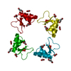 1bhpS S: Starting model for refinement |
|---|---|
| Similar structure data |


-Links
 PDBj
PDBj


- Assembly
| Deposited unit | 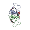
| |||||||||
|---|---|---|---|---|---|---|---|---|---|---|
| 1 |
| |||||||||
| Unit cell |
| |||||||||
| Components on special symmetry positions |
|
-Components
| #1: Protein/peptide | Mass: 4936.943 Da / Num. of mol.: 2 / Source method: isolated from a natural source / Source: (natural) #2: Chemical |   Mass: 172.202 Da / Num. of mol.: 2 / Source method: obtained synthetically / Formula: C7H8O3S Mass: 172.202 Da / Num. of mol.: 2 / Source method: obtained synthetically / Formula: C7H8O3S#3: Chemical |   Type: L-peptide linking / Mass: 105.093 Da / Num. of mol.: 2 / Source method: obtained synthetically / Formula: C3H7NO3 Type: L-peptide linking / Mass: 105.093 Da / Num. of mol.: 2 / Source method: obtained synthetically / Formula: C3H7NO3#4: Water | ChemComp-HOH / |  Mass: 18.015 Da / Num. of mol.: 92 / Source method: isolated from a natural source / Formula: H2O Mass: 18.015 Da / Num. of mol.: 92 / Source method: isolated from a natural source / Formula: H2OHas protein modification | Y | |
|---|
-Experimental details
-Experiment
| Experiment | Method: X-RAY DIFFRACTION / Number of used crystals: 1 |
|---|
- Sample preparation
| Crystal | Density Matthews: 2.83 Å3/Da / Density % sol: 56.58 % |
|---|---|
| Crystal grow | Temperature: 289 K / Method: vapor diffusion, hanging drop / pH: 5.6 Details: PEG 2000, citrate, ammonium sulfate, toluene sulfate, pH 5.6, VAPOR DIFFUSION, HANGING DROP, temperature 289K |
-Data collection
| Diffraction | Mean temperature: 298 K |
|---|---|
| Diffraction source | Source: ROTATING ANODE / Type: RIGAKU RU200 / Wavelength: 1.5418 |
| Detector | Type: ENRAF-NONIUS FAST / Detector: AREA DETECTOR / Date: Jun 21, 1994 / Details: monochromator |
| Radiation | Monochromator: graphite / Protocol: SINGLE WAVELENGTH / Monochromatic (M) / Laue (L): M / Scattering type: x-ray |
| Radiation wavelength | Wavelength: 1.5418 Å / Relative weight: 1 |
| Reflection | Resolution: 1.9→30 Å / Num. all: 8963 / Num. obs: 8963 / % possible obs: 99 % / Observed criterion σ(F): 0 / Observed criterion σ(I): 0 / Redundancy: 15.1 % / Rmerge(I) obs: 0.078 |
| Reflection shell | Resolution: 1.9→1.95 Å / Redundancy: 12.2 % / Rmerge(I) obs: 0.23 / Mean I/σ(I) obs: 8.1 / Num. unique all: 364 / % possible all: 54 |
- Processing
| Software |
| |||||||||||||||||||||||||
|---|---|---|---|---|---|---|---|---|---|---|---|---|---|---|---|---|---|---|---|---|---|---|---|---|---|---|
| Refinement | Method to determine structure: MOLECULAR REPLACEMENT Starting model: 1bhp Resolution: 1.9→30 Å / Cross valid method: THROUGHOUT / σ(F): 0 / σ(I): 0 / Stereochemistry target values: Engh & Huber
| |||||||||||||||||||||||||
| Refinement step | Cycle: LAST / Resolution: 1.9→30 Å
| |||||||||||||||||||||||||
| Refine LS restraints |
| |||||||||||||||||||||||||
| LS refinement shell | Resolution: 1.9→1.95 Å
|
