 Movie
Movie Controller

[English] 日本語
 Yorodumi
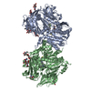
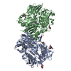
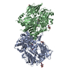
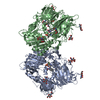
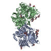
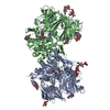
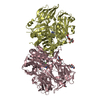
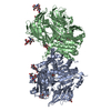
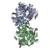
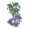
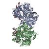
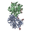
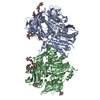
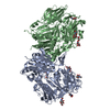
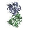
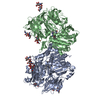
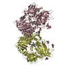
- PDB-1wcy: Crystal Structure Of Human Dipeptidyl Peptidase IV (DPPIV) Comple... -
+ Open data

- Basic information
| Entry | Database: PDB / ID: 1wcy | |||||||||
|---|---|---|---|---|---|---|---|---|---|---|
| Title | Crystal Structure Of Human Dipeptidyl Peptidase IV (DPPIV) Complex With Diprotin A | |||||||||
 Components Components |
| |||||||||
 Keywords Keywords | HYDROLASE / Serine protease / Dipeptidyl peptidase IV / CD26 / Prolyl oligopeptidase / BETA-propeller structure / Diprotin A | |||||||||
| Function / homology |  Function and homology information Function and homology informationglucagon processing / negative regulation of neutrophil chemotaxis / regulation of cell-cell adhesion mediated by integrin / Synthesis, secretion, and inactivation of Glucose-dependent Insulinotropic Polypeptide (GIP) / dipeptidyl-peptidase IV / negative regulation of extracellular matrix disassembly / chemorepellent activity / intercellular canaliculus / psychomotor behavior / dipeptidyl-peptidase activity ...glucagon processing / negative regulation of neutrophil chemotaxis / regulation of cell-cell adhesion mediated by integrin / Synthesis, secretion, and inactivation of Glucose-dependent Insulinotropic Polypeptide (GIP) / dipeptidyl-peptidase IV / negative regulation of extracellular matrix disassembly / chemorepellent activity / intercellular canaliculus / psychomotor behavior / dipeptidyl-peptidase activity / peptide hormone processing / lamellipodium membrane / locomotory exploration behavior / aminopeptidase activity / endocytic vesicle / endothelial cell migration / behavioral fear response / T cell costimulation / receptor-mediated endocytosis of virus by host cell / serine-type peptidase activity / T cell activation / Synthesis, secretion, and inactivation of Glucagon-like Peptide-1 (GLP-1) / lamellipodium / virus receptor activity / protease binding / membrane fusion / response to hypoxia / receptor-mediated virion attachment to host cell / cell adhesion / apical plasma membrane / membrane raft / signaling receptor binding / serine-type endopeptidase activity / lysosomal membrane / focal adhesion / positive regulation of cell population proliferation / symbiont entry into host cell / cell surface / protein homodimerization activity / proteolysis / extracellular exosome / extracellular region / membrane / identical protein binding / plasma membrane Similarity search - Function | |||||||||
| Biological species |  Homo sapiens (human) Homo sapiens (human) | |||||||||
| Method |  X-RAY DIFFRACTION / SYNCHROTRON / MOLECULAR REPLACEMENT / Resolution: 2.2 Å X-RAY DIFFRACTION / SYNCHROTRON / MOLECULAR REPLACEMENT / Resolution: 2.2 Å | |||||||||
 Authors Authors | Hiramatsu, H. / Yamamoto, A. / Kyono, K. / Higashiyama, Y. / Fukushima, C. / Shima, H. / Sugiyama, S. / Inaka, K. / Shimizu, R. | |||||||||
 Citation Citation | Journal: Biol.Chem. / Year: 2004 Title: The crystal structure of human dipeptidyl peptidase IV (DPPIV) complex with diprotin A Authors: Hiramatsu, H. / Yamamoto, A. / Kyono, K. / Higashiyama, Y. / Fukushima, C. / Shima, H. / Sugiyama, S. / Inaka, K. / Shimizu, R. | |||||||||
| History |
|
- Structure visualization
| Structure viewer | Molecule: MolmilJmol/JSmol |
|---|
- Downloads & links
-Download
| PDBx/mmCIF format | 1wcy.cif.gz | 338.6 KB | Display | PDBx/mmCIF format |
|---|---|---|---|---|
| PDB format | pdb1wcy.ent.gz | 271.6 KB | Display | PDB format |
| PDBx/mmJSON format | 1wcy.json.gz | Tree view | PDBx/mmJSON format | |
| Others |  Other downloads Other downloads |
-Validation report
| Arichive directory | https://data.pdbj.org/pub/pdb/validation_reports/wc/1wcyftp://data.pdbj.org/pub/pdb/validation_reports/wc/1wcy | HTTPS FTP |
|---|
-Related structure data
| Related structure data |  1j2eS S: Starting model for refinement |
|---|---|
| Similar structure data |


-Links
 PDBj
PDBj


- Assembly
| Deposited unit | 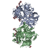
| ||||||||
|---|---|---|---|---|---|---|---|---|---|
| 1 |
| ||||||||
| Unit cell |
|
-Components
-Protein / Protein/peptide / Non-polymers , 3 types, 1241 molecules ABCD
| #1: Protein | Mass: 85880.031 Da / Num. of mol.: 2 / Fragment: residues 33-772 Source method: isolated from a genetically manipulated source Source: (gene. exp.) Homo sapiens (human) / Cell line (production host): Sf21 / Production host:   Spodoptera frugiperda (fall armyworm) / References: UniProt: P27487, dipeptidyl-peptidase IV Spodoptera frugiperda (fall armyworm) / References: UniProt: P27487, dipeptidyl-peptidase IV#2: Protein/peptide | Mass: 341.446 Da / Num. of mol.: 2 / Source method: obtained synthetically / Details: The tripeptide was chemically synthesized #6: Water | ChemComp-HOH / | Mass: 18.015 Da / Num. of mol.: 1237 / Source method: isolated from a natural source / Formula: H2O |
|---|
-Sugars , 3 types, 12 molecules 
| #3: Polysaccharide | Source method: isolated from a genetically manipulated source #4: Polysaccharide | 2-acetamido-2-deoxy-beta-D-glucopyranose-(1-4)-2-acetamido-2-deoxy-beta-D-glucopyranose Source method: isolated from a genetically manipulated source #5: Sugar | ChemComp-NAG /  Type: D-saccharide, beta linking / Mass: 221.208 Da / Num. of mol.: 5 Type: D-saccharide, beta linking / Mass: 221.208 Da / Num. of mol.: 5Source method: isolated from a genetically manipulated source Formula: C8H15NO6 |
|---|
-Details
| Has protein modification | Y |
|---|
-Experimental details
-Experiment
| Experiment | Method: X-RAY DIFFRACTION / Number of used crystals: 1 |
|---|
- Sample preparation
| Crystal | Density Matthews: 2.96 Å3/Da / Density % sol: 58.5 % |
|---|---|
| Crystal grow | Temperature: 293 K / Method: vapor diffusion, sitting drop / pH: 9.5 Details: 18% PEG4000, 0.18M Sodium acetate, 0.18M Gly-NaOH, pH 9.5, VAPOR DIFFUSION, SITTING DROP, temperature 293K |
-Data collection
| Diffraction | Mean temperature: 100 K |
|---|---|
| Diffraction source | Source: SYNCHROTRON / Site: SPring-8  / Beamline: BL32B2 / Wavelength: 1 Å / Beamline: BL32B2 / Wavelength: 1 Å |
| Detector | Type: RIGAKU RAXIS V / Detector: IMAGE PLATE |
| Radiation | Protocol: SINGLE WAVELENGTH / Monochromatic (M) / Laue (L): M / Scattering type: x-ray |
| Radiation wavelength | Wavelength: 1 Å / Relative weight: 1 |
| Reflection | Resolution: 2.2→20 Å / Num. all: 103739 / Num. obs: 103532 / % possible obs: 99.8 % / Observed criterion σ(F): 0 / Observed criterion σ(I): 0 / Redundancy: 6.9 % / Biso Wilson estimate: 20.2 Å2 / Rmerge(I) obs: 0.099 / Net I/σ(I): 5 |
| Reflection shell | Resolution: 2.2→2.32 Å / Redundancy: 6.8 % / Rmerge(I) obs: 0.332 / Mean I/σ(I) obs: 2.3 / % possible all: 99.6 |
- Processing
| Software |
| |||||||||||||||||||||||||
|---|---|---|---|---|---|---|---|---|---|---|---|---|---|---|---|---|---|---|---|---|---|---|---|---|---|---|
| Refinement | Method to determine structure: MOLECULAR REPLACEMENT Starting model: PDB ENTRY 1J2E Resolution: 2.2→20 Å / Rfactor Rfree error: 0.004 / Isotropic thermal model: RESTRAINED / Cross valid method: THROUGHOUT / σ(F): 0 / Stereochemistry target values: Engh & Huber
| |||||||||||||||||||||||||
| Displacement parameters | Biso mean: 28.8 Å2
| |||||||||||||||||||||||||
| Refine analyze |
| |||||||||||||||||||||||||
| Refinement step | Cycle: LAST / Resolution: 2.2→20 Å
| |||||||||||||||||||||||||
| Refine LS restraints |
| |||||||||||||||||||||||||
| LS refinement shell | Resolution: 2.2→2.34 Å / Rfactor Rfree error: 0.009 / Total num. of bins used: 6
|
