 Movie
Movie Controller

[English] 日本語
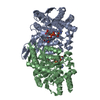
 Yorodumi
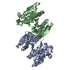
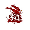
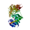
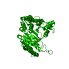
- PDB-1wal: 3-ISOPROPYLMALATE DEHYDROGENASE (IPMDH) MUTANT (M219A)FROM THERMU... -
+ Open data

- Basic information
| Entry | Database: PDB / ID: 1wal | ||||||
|---|---|---|---|---|---|---|---|
| Title | 3-ISOPROPYLMALATE DEHYDROGENASE (IPMDH) MUTANT (M219A)FROM THERMUS THERMOPHILUS | ||||||
 Components Components | PROTEIN (3-ISOPROPYLMALATE DEHYDROGENASE) | ||||||
 Keywords Keywords | OXIDOREDUCTASE / LEUCINE BIOSYNTHESIS / NAD-DEPENDANT ENZYME | ||||||
| Function / homology |  Function and homology information Function and homology information3-isopropylmalate dehydrogenase / 3-isopropylmalate dehydrogenase activity / L-leucine biosynthetic process / NAD binding / magnesium ion binding / cytosol Similarity search - Function | ||||||
| Biological species |   Thermus thermophilus (bacteria) Thermus thermophilus (bacteria) | ||||||
| Method |  X-RAY DIFFRACTION / MOLECULAR REPLACEMENT / Resolution: 2.27 Å X-RAY DIFFRACTION / MOLECULAR REPLACEMENT / Resolution: 2.27 Å | ||||||
 Authors Authors | Wallon, G. / Kryger, G. / Lovett, S.T. / Oshima, T. / Ringe, D. / Petsko, G.A. | ||||||
 Citation Citation | Journal: J.Mol.Biol. / Year: 1997 Title: Crystal structures of Escherichia coli and Salmonella typhimurium 3-isopropylmalate dehydrogenase and comparison with their thermophilic counterpart from Thermus thermophilus. Authors: Wallon, G. / Kryger, G. / Lovett, S.T. / Oshima, T. / Ringe, D. / Petsko, G.A. | ||||||
| History |
|
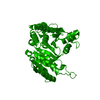
- Structure visualization
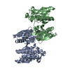
| Structure viewer | Molecule: MolmilJmol/JSmol |
|---|
- Downloads & links
-Download
| PDBx/mmCIF format | 1wal.cif.gz | 77.7 KB | Display | PDBx/mmCIF format |
|---|---|---|---|---|
| PDB format | pdb1wal.ent.gz | 58.1 KB | Display | PDB format |
| PDBx/mmJSON format | 1wal.json.gz | Tree view | PDBx/mmJSON format | |
| Others |  Other downloads Other downloads |
-Validation report
| Arichive directory | https://data.pdbj.org/pub/pdb/validation_reports/wa/1walftp://data.pdbj.org/pub/pdb/validation_reports/wa/1wal | HTTPS FTP |
|---|
-Related structure data
| Related structure data | 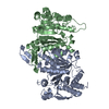 1cm7C  1cnzC  1ipdS S: Starting model for refinement C: citing same article ( |
|---|---|
| Similar structure data |
-Links
 PDBj
PDBj

- Assembly
| Deposited unit | 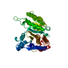
| ||||||||
|---|---|---|---|---|---|---|---|---|---|
| 1 | 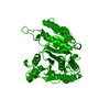
| ||||||||
| Unit cell |
|
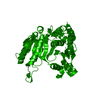
-Components
| #1: Protein | Mass: 36694.926 Da / Num. of mol.: 1 / Mutation: M219A Source method: isolated from a genetically manipulated source Source: (gene. exp.) Thermus thermophilus (bacteria) / Gene: LEUB / Plasmid: PET21C LEUB / Species (production host): Escherichia coli / Gene (production host): LEUB / Production host: |
|---|---|
| #2: Water | ChemComp-HOH /  Mass: 18.015 Da / Num. of mol.: 97 / Source method: isolated from a natural source / Formula: H2O Mass: 18.015 Da / Num. of mol.: 97 / Source method: isolated from a natural source / Formula: H2O |
-Experimental details
-Experiment
| Experiment | Method: X-RAY DIFFRACTION |
|---|
- Sample preparation
| Crystal | Density Matthews: 2.7 Å3/Da / Density % sol: 67.82 % | ||||||||||||||||||||||||
|---|---|---|---|---|---|---|---|---|---|---|---|---|---|---|---|---|---|---|---|---|---|---|---|---|---|
| Crystal grow | pH: 7.6 / Details: 6 MG/ML PROTEIN IN 1.4 M AMMONIUM SULFATE, pH 7.6 | ||||||||||||||||||||||||
| Crystal grow | *PLUS Method: vapor diffusion, hanging drop | ||||||||||||||||||||||||
| Components of the solutions | *PLUS
|
-Data collection
| Diffraction | Mean temperature: 277 K |
|---|---|
| Diffraction source | Source: ROTATING ANODE / Type: RIGAKU RU200 / Wavelength: 1.5418 |
| Detector | Type: RIGAKU RAXIS IIC / Detector: IMAGE PLATE / Details: COLLIMATOR |
| Radiation | Monochromator: GRAPHITE CRYSTAL / Protocol: SINGLE WAVELENGTH / Monochromatic (M) / Laue (L): M / Scattering type: x-ray |
| Radiation wavelength | Wavelength: 1.5418 Å / Relative weight: 1 |
| Reflection | Resolution: 2.27→30 Å / Num. obs: 54255 / % possible obs: 84 % / Observed criterion σ(I): 1 / Redundancy: 2.4 % / Rsym value: 0.055 |
| Reflection | *PLUS Num. obs: 22587 / Num. measured all: 54255 / Rmerge(I) obs: 0.055 |
- Processing
| Software |
| ||||||||||||||||||||||||||||||||||||||||||||||||||||||||||||
|---|---|---|---|---|---|---|---|---|---|---|---|---|---|---|---|---|---|---|---|---|---|---|---|---|---|---|---|---|---|---|---|---|---|---|---|---|---|---|---|---|---|---|---|---|---|---|---|---|---|---|---|---|---|---|---|---|---|---|---|---|---|
| Refinement | Method to determine structure: MOLECULAR REPLACEMENT Starting model: 1IPD Resolution: 2.27→6 Å / σ(F): 1
| ||||||||||||||||||||||||||||||||||||||||||||||||||||||||||||
| Displacement parameters | Biso mean: 24 Å2 | ||||||||||||||||||||||||||||||||||||||||||||||||||||||||||||
| Refinement step | Cycle: LAST / Resolution: 2.27→6 Å
| ||||||||||||||||||||||||||||||||||||||||||||||||||||||||||||
| Refine LS restraints |
| ||||||||||||||||||||||||||||||||||||||||||||||||||||||||||||
| Software | *PLUS Name: X-PLOR / Classification: refinement | ||||||||||||||||||||||||||||||||||||||||||||||||||||||||||||
| Refinement | *PLUS Rfactor obs: 0.184 | ||||||||||||||||||||||||||||||||||||||||||||||||||||||||||||
| Solvent computation | *PLUS | ||||||||||||||||||||||||||||||||||||||||||||||||||||||||||||
| Displacement parameters | *PLUS Biso mean: 24 Å2 | ||||||||||||||||||||||||||||||||||||||||||||||||||||||||||||
| Refine LS restraints | *PLUS Type: x_bond_d / Dev ideal: 0.01 |
