 Movie
Movie Controller

[English] 日本語
 Yorodumi
- PDB-1w1x: Structure of Neuraminidase from English duck subtype N6 complexed... -
+ Open data

- Basic information
| Entry | Database: PDB / ID: 1w1x | |||||||||
|---|---|---|---|---|---|---|---|---|---|---|
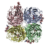
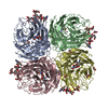
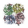
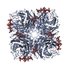
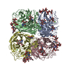
| Title | Structure of Neuraminidase from English duck subtype N6 complexed with 30 mM sialic acid (NANA, Neu5Ac), crystal soaked for 3 hours at 277 K. | |||||||||
 Components Components | NEURAMINIDASE | |||||||||
 Keywords Keywords | HYDROLASE / INFLUENZA TYPE A / NEURAMINIDASE / HB SITE / SIALIC ACID / SUBTYPE N6 | |||||||||
| Function / homology |  Function and homology information Function and homology informationexo-alpha-sialidase / exo-alpha-sialidase activity / viral budding from plasma membrane / carbohydrate metabolic process / host cell plasma membrane / virion membrane / membrane / metal ion binding Similarity search - Function | |||||||||
| Biological species |   INFLUENZA A VIRUS INFLUENZA A VIRUS | |||||||||
| Method |  X-RAY DIFFRACTION / SYNCHROTRON / MOLECULAR REPLACEMENT / Resolution: 2 Å X-RAY DIFFRACTION / SYNCHROTRON / MOLECULAR REPLACEMENT / Resolution: 2 Å | |||||||||
 Authors Authors | Rudino-Pinera, E. / Tunnah, P. / Crennell, S.J. / Webster, R.G. / Laver, W.G. / Garman, E.F. | |||||||||
 Citation Citation | Journal: To be Published Title: The Crystal Structure of Type a Influenza Virus Neuraminidase of the N6 Subtype Reveals the Existence of Two Separate Neu5Ac Binding Sites Authors: Rudino-Pinera, E. / Tunnah, P. / Crennell, S.J. / Webster, R.G. / Laver, W.G. / Garman, E.F. | |||||||||
| History |
|
- Structure visualization
| Structure viewer | Molecule: MolmilJmol/JSmol |
|---|
- Downloads & links
-Download
| PDBx/mmCIF format | 1w1x.cif.gz | 362.4 KB | Display | PDBx/mmCIF format |
|---|---|---|---|---|
| PDB format | pdb1w1x.ent.gz | 288.8 KB | Display | PDB format |
| PDBx/mmJSON format | 1w1x.json.gz | Tree view | PDBx/mmJSON format | |
| Others |  Other downloads Other downloads |
-Validation report
| Arichive directory | https://data.pdbj.org/pub/pdb/validation_reports/w1/1w1xftp://data.pdbj.org/pub/pdb/validation_reports/w1/1w1x | HTTPS FTP |
|---|
-Related structure data
| Related structure data |  1w20C  1w21C  2cmlC 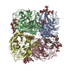 1v0zS S: Starting model for refinement C: citing same article ( |
|---|---|
| Similar structure data |

-Links
 PDBj
PDBj


- Assembly
| Deposited unit | 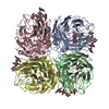
| ||||||||
|---|---|---|---|---|---|---|---|---|---|
| 1 |
| ||||||||
| Unit cell |
|
-Components
-Protein , 1 types, 4 molecules ABCD
| #1: Protein | Mass: 43011.531 Da / Num. of mol.: 4 Source method: isolated from a genetically manipulated source Details: COMPLEXED WITH SIALIC ACID / Source: (gene. exp.) INFLUENZA A VIRUSDescription: INFLUENZA VIRUS GROWN IN EMBRIONATED CHICKEN EGGS Production host:  |
|---|
-Sugars , 9 types, 25 molecules 


| #2: Polysaccharide | alpha-D-mannopyranose-(1-6)-alpha-D-mannopyranose-(1-6)-beta-D-mannopyranose Source method: isolated from a genetically manipulated source | ||||||||||||||
|---|---|---|---|---|---|---|---|---|---|---|---|---|---|---|---|
| #3: Polysaccharide | Source method: isolated from a genetically manipulated source #4: Polysaccharide | beta-D-mannopyranose-(1-4)-2-acetamido-2-deoxy-beta-D-glucopyranose-(1-4)-2-acetamido-2-deoxy-beta- ...beta-D-mannopyranose-(1-4)-2-acetamido-2-deoxy-beta-D-glucopyranose-(1-4)-2-acetamido-2-deoxy-beta-D-glucopyranose | Source method: isolated from a genetically manipulated source #5: Polysaccharide | alpha-D-mannopyranose-(1-6)-alpha-D-mannopyranose / 6alpha-alpha-mannobiose |   Source method: isolated from a genetically manipulated source Details: oligosaccharide / References: 6alpha-alpha-mannobiose #6: Polysaccharide | alpha-D-mannopyranose-(1-6)-beta-D-mannopyranose-(1-4)-2-acetamido-2-deoxy-beta-D-glucopyranose | Source method: isolated from a genetically manipulated source #7: Sugar | ChemComp-SIA /  Type: D-saccharide, alpha linking / Mass: 309.270 Da / Num. of mol.: 8 Type: D-saccharide, alpha linking / Mass: 309.270 Da / Num. of mol.: 8Source method: isolated from a genetically manipulated source Formula: C11H19NO9 #9: Sugar | ChemComp-MAN /  Type: D-saccharide, alpha linking / Mass: 180.156 Da / Num. of mol.: 5 Type: D-saccharide, alpha linking / Mass: 180.156 Da / Num. of mol.: 5Source method: isolated from a genetically manipulated source Formula: C6H12O6 #10: Sugar | ChemComp-NAG /  Type: D-saccharide, beta linking / Mass: 221.208 Da / Num. of mol.: 5 Type: D-saccharide, beta linking / Mass: 221.208 Da / Num. of mol.: 5Source method: isolated from a genetically manipulated source Formula: C8H15NO6 #13: Sugar | ChemComp-BMA / |  Type: D-saccharide, beta linking / Mass: 180.156 Da / Num. of mol.: 1 Type: D-saccharide, beta linking / Mass: 180.156 Da / Num. of mol.: 1Source method: isolated from a genetically manipulated source Formula: C6H12O6 |
-Non-polymers , 4 types, 1574 molecules 


| #8: Chemical | ChemComp-CA /  Mass: 40.078 Da / Num. of mol.: 4 / Source method: obtained synthetically / Formula: Ca Mass: 40.078 Da / Num. of mol.: 4 / Source method: obtained synthetically / Formula: Ca#11: Chemical | ChemComp-GOL /  Mass: 92.094 Da / Num. of mol.: 4 / Source method: obtained synthetically / Formula: C3H8O3 Mass: 92.094 Da / Num. of mol.: 4 / Source method: obtained synthetically / Formula: C3H8O3#12: Chemical | ChemComp-PEG / |  Mass: 106.120 Da / Num. of mol.: 1 / Source method: obtained synthetically / Formula: C4H10O3 Mass: 106.120 Da / Num. of mol.: 1 / Source method: obtained synthetically / Formula: C4H10O3#14: Water | ChemComp-HOH / | Mass: 18.015 Da / Num. of mol.: 1565 / Source method: isolated from a natural source / Formula: H2O |
|---|
-Details
| Has protein modification | Y |
|---|
-Experimental details
-Experiment
| Experiment | Method: X-RAY DIFFRACTION / Number of used crystals: 1 |
|---|
- Sample preparation
| Crystal | Density Matthews: 2.5 Å3/Da / Density % sol: 50.86 % |
|---|---|
| Crystal grow | Temperature: 293 K / pH: 7 / Details: 0.15 M NACL, 20% PEG 3350 AT 293 K, pH 7.00 |
-Data collection
| Diffraction | Mean temperature: 100 K |
|---|---|
| Diffraction source | Source: SYNCHROTRON / Site: SRS  / Beamline: PX14.2 / Wavelength: 0.9853 / Beamline: PX14.2 / Wavelength: 0.9853 |
| Detector | Type: ADSC CCD / Detector: CCD / Date: Mar 15, 2004 / Details: 1.2 METRE LONG SILICON SUBSTRATE, RHODIUM COATED |
| Radiation | Monochromator: SILICON 11 / Protocol: SINGLE WAVELENGTH / Monochromatic (M) / Laue (L): M / Scattering type: x-ray |
| Radiation wavelength | Wavelength: 0.9853 Å / Relative weight: 1 |
| Reflection | Resolution: 2→33.78 Å / Num. obs: 112032 / % possible obs: 99.4 % / Observed criterion σ(I): 0 / Redundancy: 3.3 % / Biso Wilson estimate: 16.63 Å2 / Rmerge(I) obs: 0.12 / Net I/σ(I): 4.6 |
| Reflection shell | Resolution: 2→2.11 Å / Redundancy: 3.3 % / Rmerge(I) obs: 0.3 / Mean I/σ(I) obs: 2.3 / % possible all: 99 |
- Processing
| Software |
| ||||||||||||||||||||||||||||||||||||||||||||||||||||||||||||||||||||||||||||||||||||||||||||||||||||||||||||||||||||||||||||||||||||||||||||||||||||||||||||||||||||||||||||||||||||||
|---|---|---|---|---|---|---|---|---|---|---|---|---|---|---|---|---|---|---|---|---|---|---|---|---|---|---|---|---|---|---|---|---|---|---|---|---|---|---|---|---|---|---|---|---|---|---|---|---|---|---|---|---|---|---|---|---|---|---|---|---|---|---|---|---|---|---|---|---|---|---|---|---|---|---|---|---|---|---|---|---|---|---|---|---|---|---|---|---|---|---|---|---|---|---|---|---|---|---|---|---|---|---|---|---|---|---|---|---|---|---|---|---|---|---|---|---|---|---|---|---|---|---|---|---|---|---|---|---|---|---|---|---|---|---|---|---|---|---|---|---|---|---|---|---|---|---|---|---|---|---|---|---|---|---|---|---|---|---|---|---|---|---|---|---|---|---|---|---|---|---|---|---|---|---|---|---|---|---|---|---|---|---|---|
| Refinement | Method to determine structure: MOLECULAR REPLACEMENT Starting model: PDB ENTRY 1V0Z Resolution: 2→33.71 Å / Cor.coef. Fo:Fc: 0.962 / Cor.coef. Fo:Fc free: 0.943 / SU B: 4.684 / SU ML: 0.12 / Cross valid method: THROUGHOUT / ESU R: 0.178 / ESU R Free: 0.154 / Stereochemistry target values: MAXIMUM LIKELIHOOD Details: UNLIKE OTHER VIRAL NEURAMINIDASES SOLVED TO DATE, N6 HAS A FULL TETRAMER IN THE ASYMMETRIC UNIT
| ||||||||||||||||||||||||||||||||||||||||||||||||||||||||||||||||||||||||||||||||||||||||||||||||||||||||||||||||||||||||||||||||||||||||||||||||||||||||||||||||||||||||||||||||||||||
| Solvent computation | Ion probe radii: 0.8 Å / Shrinkage radii: 0.8 Å / VDW probe radii: 1.4 Å / Solvent model: BABINET MODEL WITH MASK | ||||||||||||||||||||||||||||||||||||||||||||||||||||||||||||||||||||||||||||||||||||||||||||||||||||||||||||||||||||||||||||||||||||||||||||||||||||||||||||||||||||||||||||||||||||||
| Displacement parameters | Biso mean: 21.28 Å2
| ||||||||||||||||||||||||||||||||||||||||||||||||||||||||||||||||||||||||||||||||||||||||||||||||||||||||||||||||||||||||||||||||||||||||||||||||||||||||||||||||||||||||||||||||||||||
| Refinement step | Cycle: LAST / Resolution: 2→33.71 Å
| ||||||||||||||||||||||||||||||||||||||||||||||||||||||||||||||||||||||||||||||||||||||||||||||||||||||||||||||||||||||||||||||||||||||||||||||||||||||||||||||||||||||||||||||||||||||
| Refine LS restraints |
|