Resolution: 2.5→28.66 Å / Num. obs: 49807 / % possible obs: 98.6 % / Redundancy: 4.6 % / Biso Wilson estimate: 82.46 Å2 / Rmerge(I) obs: 0.067 / Net I/σ(I): 13.7
Reflection shell
Resolution: 2.5→2.64 Å / Redundancy: 3 % / Rmerge(I) obs: 0.727 / Mean I/σ(I) obs: 2.3 / Num. unique all: 6756 / % possible all: 93.3
-
Processing
Software
Name
Version
Classification
XDS
datascaling
SCALA
datascaling
SHELXD
+ AutoSHARP
phasing
REFMAC
5.2.0005
refinement
XDS
datareduction
CCP4
(SCALA)
datascaling
autoSHARP
phasing
Refinement
Method to determine structure: MAD / Resolution: 2.6→28.66 Å / Cor.coef. Fo:Fc: 0.941 / Cor.coef. Fo:Fc free: 0.917 / SU B: 29.814 / SU ML: 0.286 / TLS residual ADP flag: LIKELY RESIDUAL / Cross valid method: THROUGHOUT / ESU R: 0.701 / ESU R Free: 0.333 Stereochemistry target values: MAXIMUM LIKELIHOOD WITH PHASES Details: 1.HYDROGENS HAVE BEEN ADDED IN THE RIDING POSITIONS. 2.THE DENSITIES FOR THE FOLLOWING REGIONS, 66-96, 178-185, 268-275, 138-142, ARE VERY POOR. ALTHOUGH MODEL WAS BUILT FOR SOME OF THESE ...Details: 1.HYDROGENS HAVE BEEN ADDED IN THE RIDING POSITIONS. 2.THE DENSITIES FOR THE FOLLOWING REGIONS, 66-96, 178-185, 268-275, 138-142, ARE VERY POOR. ALTHOUGH MODEL WAS BUILT FOR SOME OF THESE REGIONS, THEY MAY CONTAIN ERRORS. 3.A FERRIC IRON IS MODELLED FOR EACH CHAIN. THIS ASSIGNMENT WAS BASED ON HOMOLOGS (1IZ3 AND 1MZE). HOWEVER, THIS CRYSTAL WAS OBTAINED IN BUFFER CONTAINING COPPER. THERE IS NO EVIDENCE TO EXCLUED THE POSSIBILITY THE ION BEING OTHER METALS SUCH AS COPPER. 4.D278-D285 IS A PART OF HELIX, THE REFINEMENT OF THIS REGION IS NOT STABLE DUE TO THE POOR DENSITY.
Rfactor
Num. reflection
% reflection
Selection details
Rfree
0.27227
2243
5 %
RANDOM
Rwork
0.22144
-
-
-
obs
0.22399
42483
99.3 %
-
Solvent computation
Ion probe radii: 0.8 Å / Shrinkage radii: 0.8 Å / VDW probe radii: 1.2 Å / Solvent model: BABINET MODEL WITH MASK
In the structure databanks used in Yorodumi, some data are registered as the other names, "COVID-19 virus" and "2019-nCoV". Here are the details of the virus and the list of structure data.
Jan 31, 2019. EMDB accession codes are about to change! (news from PDBe EMDB page)
EMDB accession codes are about to change! (news from PDBe EMDB page)
The allocation of 4 digits for EMDB accession codes will soon come to an end. Whilst these codes will remain in use, new EMDB accession codes will include an additional digit and will expand incrementally as the available range of codes is exhausted. The current 4-digit format prefixed with “EMD-” (i.e. EMD-XXXX) will advance to a 5-digit format (i.e. EMD-XXXXX), and so on. It is currently estimated that the 4-digit codes will be depleted around Spring 2019, at which point the 5-digit format will come into force.
The EM Navigator/Yorodumi systems omit the EMD- prefix.
Related info.:Q: What is EMD? / ID/Accession-code notation in Yorodumi/EM Navigator
Yorodumi is a browser for structure data from EMDB, PDB, SASBDB, etc.
This page is also the successor to EM Navigator detail page, and also detail information page/front-end page for Omokage search.
The word "yorodu" (or yorozu) is an old Japanese word meaning "ten thousand". "mi" (miru) is to see.
Related info.:EMDB / PDB / SASBDB / Comparison of 3 databanks / Yorodumi Search / Aug 31, 2016. New EM Navigator & Yorodumi / Yorodumi Papers / Jmol/JSmol / Function and homology information / Changes in new EM Navigator and Yorodumi
 Movie
Movie Controller

 Yorodumi
 Open data
 Basic information
 Components
 Keywords
 Function and homology information
 X-RAY DIFFRACTION /
  Authors
 Citation
 Structure visualization
 Downloads & links
 Other downloads
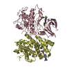

 PDBj
PDBj
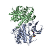
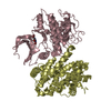
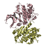
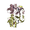
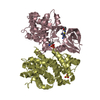
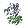
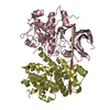
 Assembly
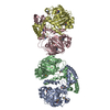

 Mass: 55.845 Da / Num. of mol.: 4 / Source method: obtained synthetically / Formula: Fe
 Mass: 18.015 Da / Num. of mol.: 22 / Source method: isolated from a natural source / Formula: H2O
 Sample preparation
 Processing