Monochromator: Double Crystal Si(111) / Protocol: SINGLE WAVELENGTH / Monochromatic (M) / Laue (L): M / Scattering type: x-ray
Radiation wavelength
Wavelength: 0.979594 Å / Relative weight: 1
Reflection
Resolution: 1.2→55.24 Å / Num. obs: 88939 / % possible obs: 99 % / Redundancy: 3 % / Rmerge(I) obs: 0.055 / Rsym value: 0.055 / Net I/σ(I): 7.1
Reflection shell
Diffraction-ID: 1
Resolution (Å)
% possible obs (%)
Redundancy (%)
Rmerge(I) obs
Mean I/σ(I) obs
Num. measured obs
Rsym value
1.2-1.23
93.5
1.9
0.575
1.2
6237
0.575
1.23-1.26
96.8
2.3
0.497
1.2
6264
0.497
1.26-1.3
98.5
2.5
0.391
1.8
6209
0.391
1.3-1.34
99.7
2.6
0.335
2.2
6082
0.335
1.34-1.39
99.8
2.6
0.249
2.8
5963
0.249
1.39-1.43
99.6
2.6
0.192
3.8
5639
0.192
1.43-1.49
99.7
2.7
0.154
2.9
5515
0.154
1.49-1.55
99.7
2.7
0.127
4.3
5326
0.127
1.55-1.62
99.7
2.7
0.111
5.7
5087
0.111
1.62-1.7
99.9
2.7
0.101
6.3
4884
0.101
1.7-1.79
99.9
2.7
0.093
6.6
4620
0.093
1.79-1.9
99.9
2.7
0.088
6.3
4379
0.088
1.9-2.03
100
2.9
0.087
6.9
4151
0.087
2.03-2.19
100
3.4
0.077
8.3
3820
0.077
2.19-2.4
100
4.1
0.067
9.7
3541
0.067
2.4-2.68
100
5.2
0.067
9.2
3183
0.067
2.68-3.1
100
5.5
0.055
11.3
2829
0.055
3.1-3.79
100
5.5
0.046
12.9
2376
0.046
3.79-5.37
100
5.1
0.042
13.4
1823
0.042
5.37-55.24
99.9
4.8
0.042
12.8
1011
0.042
-
Phasing
Phasing
Method: molecular replacement
-
Processing
Software
Name
Version
Classification
NB
SHELX
refinement
SCALA
datascaling
MOSFLM
datareduction
CCP4
(SCALA)
datascaling
CNS
phasing
SHELXL-97
refinement
Refinement
Resolution: 1.2→55 Å / Num. parameters: 19113 / Num. restraintsaints: 24162 / Cross valid method: FREE R / σ(F): 0 / Stereochemistry target values: ENGH AND HUBER Details: 1. THE CRYSTAL WAS TWINNED ACCORDING TO THE TWIN LAW 1 0 0 -1 -1 0 0 0 -1. THE TWINNING FRACTION WAS REFINED IN SHELXL AND WAS 0.31 IN THE FINAL CYCLE. 2. THE FINAL REFINEMENT CYCLE WAS ...Details: 1. THE CRYSTAL WAS TWINNED ACCORDING TO THE TWIN LAW 1 0 0 -1 -1 0 0 0 -1. THE TWINNING FRACTION WAS REFINED IN SHELXL AND WAS 0.31 IN THE FINAL CYCLE. 2. THE FINAL REFINEMENT CYCLE WAS AGAINST ALL DATA. THE FREE R VALUES ABOVE ARE FROM THE PREVIOUS CYCLE. 3. THE TWIN LAW WAS USED IN SELECTING THE REFLECTIONS FOR THE TEST SET.
In the structure databanks used in Yorodumi, some data are registered as the other names, "COVID-19 virus" and "2019-nCoV". Here are the details of the virus and the list of structure data.
Jan 31, 2019. EMDB accession codes are about to change! (news from PDBe EMDB page)
EMDB accession codes are about to change! (news from PDBe EMDB page)
The allocation of 4 digits for EMDB accession codes will soon come to an end. Whilst these codes will remain in use, new EMDB accession codes will include an additional digit and will expand incrementally as the available range of codes is exhausted. The current 4-digit format prefixed with “EMD-” (i.e. EMD-XXXX) will advance to a 5-digit format (i.e. EMD-XXXXX), and so on. It is currently estimated that the 4-digit codes will be depleted around Spring 2019, at which point the 5-digit format will come into force.
The EM Navigator/Yorodumi systems omit the EMD- prefix.
Related info.:Q: What is EMD? / ID/Accession-code notation in Yorodumi/EM Navigator
Yorodumi is a browser for structure data from EMDB, PDB, SASBDB, etc.
This page is also the successor to EM Navigator detail page, and also detail information page/front-end page for Omokage search.
The word "yorodu" (or yorozu) is an old Japanese word meaning "ten thousand". "mi" (miru) is to see.
Related info.:EMDB / PDB / SASBDB / Comparison of 3 databanks / Yorodumi Search / Aug 31, 2016. New EM Navigator & Yorodumi / Yorodumi Papers / Jmol/JSmol / Function and homology information / Changes in new EM Navigator and Yorodumi
 Movie
Movie Controller

 Yorodumi
 Open data
 Basic information
 Components
 Keywords
 Function and homology information
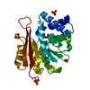
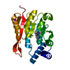
 Thermotoga maritima (bacteria)
 X-RAY DIFFRACTION /
  Authors
 Citation
 Structure visualization
 Downloads & links
 Other downloads


 PDBj
PDBj Assembly

 Mass: 62.068 Da / Num. of mol.: 1 / Source method: obtained synthetically / Formula: C2H6O2
 Mass: 18.015 Da / Num. of mol.: 153 / Source method: isolated from a natural source / Formula: H2O
 Sample preparation
 / Beamline: 8.3.1 / Wavelength: 0.979594
  Processing