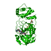
SEQUENCE CLONING ARTIFACT: THE CONSTRUCT WAS PCR AMPLIFIED WITH TAQ POLYMERASE. SEQUENCING OF THE ... SEQUENCE CLONING ARTIFACT: THE CONSTRUCT WAS PCR AMPLIFIED WITH TAQ POLYMERASE. SEQUENCING OF THE CLONED CONSTRUCT INDICATED THAT ARG IN POSITION 33 WAS MUTATED TO ALA. HOWEVER, THE LOCATION OF ALA33 WITHIN THE STRUCTURE SUGGESTS THAT A LARGE SIDE CHAIN AT THIS POSITION IS UNLIKELY WITHOUT SIGNIFICANT STRUCTURAL CHANGES.
Monochromator: Double Crystal Si(111) / Protocol: SINGLE WAVELENGTH / Monochromatic (M) / Laue (L): M / Scattering type: x-ray
Radiation wavelength
Wavelength: 1.11587 Å / Relative weight: 1
Reflection
Resolution: 2.2→63.66 Å / Num. obs: 26842 / % possible obs: 100 % / Redundancy: 7.4 % / Biso Wilson estimate: 53.64 Å2 / Rsym value: 0.068 / Net I/σ(I): 13.9
Reflection shell
Resolution: 2.2→2.32 Å / Redundancy: 4.7 % / Mean I/σ(I) obs: 2.3 / Num. unique all: 3836 / Rsym value: 0.588 / % possible all: 100
-
Processing
Software
Name
Version
Classification
REFMAC
5.2.0001
refinement
SCALA
4.2)
datascaling
CNS
refinement
MOSFLM
datareduction
CCP4
(SCALA)
datascaling
CNS
phasing
Refinement
Method to determine structure: MOLECULAR REPLACEMENT / Resolution: 2.2→63.66 Å / Cor.coef. Fo:Fc: 0.959 / Cor.coef. Fo:Fc free: 0.936 / SU B: 8.866 / SU ML: 0.116 / TLS residual ADP flag: LIKELY RESIDUAL / Cross valid method: THROUGHOUT / ESU R: 0.152 / ESU R Free: 0.144 / Stereochemistry target values: MAXIMUM LIKELIHOOD Details: THE METAL SITE COORDINATED BY CYS98, CYS101, CYS116, CYS119 IS TENATIVELY ASSIGNED AS ZINC BASED ON SEQUENCE HOMOLOGY. IT IS CONSISTENT WITH ELECTRON DENSITY AND COORDINATION. OTHER METALS ...Details: THE METAL SITE COORDINATED BY CYS98, CYS101, CYS116, CYS119 IS TENATIVELY ASSIGNED AS ZINC BASED ON SEQUENCE HOMOLOGY. IT IS CONSISTENT WITH ELECTRON DENSITY AND COORDINATION. OTHER METALS SUCH AS IRON ARE ALSO POSSIBLE. ELECTRON DENSITY AT THE C-TERMINUS IS POOR.
Rfactor
Num. reflection
% reflection
Selection details
Rfree
0.21955
1371
5.1 %
RANDOM
Rwork
0.18668
-
-
-
obs
0.18831
25436
99.87 %
-
Solvent computation
Ion probe radii: 0.8 Å / Shrinkage radii: 0.8 Å / VDW probe radii: 1.2 Å / Solvent model: BABINET MODEL WITH MASK
Displacement parameters
Biso mean: 68.881 Å2
Baniso -1
Baniso -2
Baniso -3
1-
1.85 Å2
0.92 Å2
0 Å2
2-
-
1.85 Å2
0 Å2
3-
-
-
-2.77 Å2
Refinement step
Cycle: LAST / Resolution: 2.2→63.66 Å
Protein
Nucleic acid
Ligand
Solvent
Total
Num. atoms
2053
0
25
96
2174
Refine LS restraints
Refine-ID
Type
Dev ideal
Dev ideal target
Number
X-RAY DIFFRACTION
r_bond_refined_d
0.017
0.021
2149
X-RAY DIFFRACTION
r_angle_refined_deg
1.509
1.936
2925
X-RAY DIFFRACTION
r_dihedral_angle_1_deg
5.824
5
256
X-RAY DIFFRACTION
r_dihedral_angle_2_deg
34.71
23.524
105
X-RAY DIFFRACTION
r_dihedral_angle_3_deg
14.851
15
351
X-RAY DIFFRACTION
r_dihedral_angle_4_deg
15.135
15
16
X-RAY DIFFRACTION
r_chiral_restr
0.102
0.2
312
X-RAY DIFFRACTION
r_gen_planes_refined
0.006
0.02
1646
X-RAY DIFFRACTION
r_nbd_refined
0.195
0.2
890
X-RAY DIFFRACTION
r_xyhbond_nbd_refined
0.137
0.2
113
X-RAY DIFFRACTION
r_symmetry_vdw_refined
0.201
0.2
62
X-RAY DIFFRACTION
r_symmetry_hbond_refined
0.178
0.2
15
X-RAY DIFFRACTION
r_mcbond_it
2.452
3
1324
X-RAY DIFFRACTION
r_mcangle_it
3.466
5
2074
X-RAY DIFFRACTION
r_scbond_it
6.251
8
963
X-RAY DIFFRACTION
r_scangle_it
7.928
11
851
LS refinement shell
Resolution: 2.2→2.257 Å / Total num. of bins used: 20
Rfactor
Num. reflection
% reflection
Rfree
0.298
113
5.85 %
Rwork
0.25
1818
-
Refinement TLS params.
Method: refined / Refine-ID: X-RAY DIFFRACTION
ID
L11 (°2)
L12 (°2)
L13 (°2)
L22 (°2)
L23 (°2)
L33 (°2)
S11 (Å °)
S12 (Å °)
S13 (Å °)
S21 (Å °)
S22 (Å °)
S23 (Å °)
S31 (Å °)
S32 (Å °)
S33 (Å °)
T11 (Å2)
T12 (Å2)
T13 (Å2)
T22 (Å2)
T23 (Å2)
T33 (Å2)
Origin x (Å)
Origin y (Å)
Origin z (Å)
1
1.6906
0.7142
1.299
0.6809
1.334
5.579
0.0391
-0.2451
0.0352
0.204
-0.1411
0.0064
0.4056
0.2978
0.1021
-0.1934
-0.1513
-0.0191
-0.025
0.0859
-0.1666
82.035
41.617
8.911
2
2.2156
0.7016
1.6064
2.1159
1.2102
7.2225
-0.3726
0.0564
0.7209
-0.024
-0.1053
0.2295
-1.6316
0.2241
0.478
0.1509
-0.2671
-0.1763
-0.0264
0.1235
0.0588
93.341
68.942
-1.053
Refinement TLS group
Refine-ID: X-RAY DIFFRACTION / Selection: ALL / Auth asym-ID: A / Label asym-ID: A
ID
Refine TLS-ID
Auth seq-ID
Label seq-ID
1
1
0 - 127
12 - 139
2
2
128 - 256
140 - 268
+
About Yorodumi
-
News
-
Feb 9, 2022. New format data for meta-information of EMDB entries
New format data for meta-information of EMDB entries
Version 3 of the EMDB header file is now the official format.
The previous official version 1.9 will be removed from the archive.
In the structure databanks used in Yorodumi, some data are registered as the other names, "COVID-19 virus" and "2019-nCoV". Here are the details of the virus and the list of structure data.
Jan 31, 2019. EMDB accession codes are about to change! (news from PDBe EMDB page)
EMDB accession codes are about to change! (news from PDBe EMDB page)
The allocation of 4 digits for EMDB accession codes will soon come to an end. Whilst these codes will remain in use, new EMDB accession codes will include an additional digit and will expand incrementally as the available range of codes is exhausted. The current 4-digit format prefixed with “EMD-” (i.e. EMD-XXXX) will advance to a 5-digit format (i.e. EMD-XXXXX), and so on. It is currently estimated that the 4-digit codes will be depleted around Spring 2019, at which point the 5-digit format will come into force.
The EM Navigator/Yorodumi systems omit the EMD- prefix.
Related info.:Q: What is EMD? / ID/Accession-code notation in Yorodumi/EM Navigator
Yorodumi is a browser for structure data from EMDB, PDB, SASBDB, etc.
This page is also the successor to EM Navigator detail page, and also detail information page/front-end page for Omokage search.
The word "yorodu" (or yorozu) is an old Japanese word meaning "ten thousand". "mi" (miru) is to see.
Related info.:EMDB / PDB / SASBDB / Comparison of 3 databanks / Yorodumi Search / Aug 31, 2016. New EM Navigator & Yorodumi / Yorodumi Papers / Jmol/JSmol / Function and homology information / Changes in new EM Navigator and Yorodumi
 Movie
Movie Controller

 Yorodumi
 Open data
 Basic information
 Components
 Keywords
 Function and homology information
 X-RAY DIFFRACTION /
  Authors
 Citation
 Structure visualization
 Downloads & links
 Other downloads
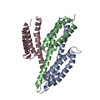

 PDBj
PDBj

 Assembly
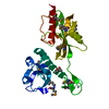

 Mass: 65.409 Da / Num. of mol.: 1 / Source method: obtained synthetically / Formula: Zn
 Mass: 118.174 Da / Num. of mol.: 3 / Source method: obtained synthetically / Formula: C6H14O2 / Comment: precipitant*YM
 Mass: 18.015 Da / Num. of mol.: 96 / Source method: isolated from a natural source / Formula: H2O
 Sample preparation
 / Beamline: 8.3.1 / Wavelength: 1.11587
  Processing