 Movie
Movie Controller

[English] 日本語
 Yorodumi
- PDB-1vbn: Escherichia coli tyrosyl-tRNA synthetase mutant complexed with Tyr-AMS -
+ Open data

- Basic information
| Entry | Database: PDB / ID: 1vbn | ||||||
|---|---|---|---|---|---|---|---|
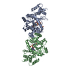
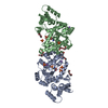
| Title | Escherichia coli tyrosyl-tRNA synthetase mutant complexed with Tyr-AMS | ||||||
 Components Components | Tyrosyl-tRNA synthetase | ||||||
 Keywords Keywords | LIGASE / Structural Genomics / RIKEN Structural Genomics/Proteomics Initiative / RSGI | ||||||
| Function / homology |  Function and homology information Function and homology informationtRNA aminoacylation / tyrosyl-tRNA aminoacylation / tyrosine-tRNA ligase / tyrosine-tRNA ligase activity / protein homodimerization activity / RNA binding / ATP binding / membrane / cytosol Similarity search - Function | ||||||
| Biological species |  | ||||||
| Method |  X-RAY DIFFRACTION / SYNCHROTRON / MOLECULAR REPLACEMENT / Resolution: 2.7 Å X-RAY DIFFRACTION / SYNCHROTRON / MOLECULAR REPLACEMENT / Resolution: 2.7 Å | ||||||
 Authors Authors | Kobayashi, T. / Sakamoto, K. / Takimura, T. / Kamata, K. / Sekine, R. / Nishimura, S. / Yokoyama, S. / RIKEN Structural Genomics/Proteomics Initiative (RSGI) | ||||||
 Citation Citation | Journal: Proc.Natl.Acad.Sci.USA / Year: 2005 Title: Structural basis of nonnatural amino acid recognition by an engineered aminoacyl-tRNA synthetase for genetic code expansion Authors: Kobayashi, T. / Sakamoto, K. / Takimura, T. / Sekine, R. / Vincent, K. / Kamata, K. / Nishimura, S. / Yokoyama, S. | ||||||
| History |
|
- Structure visualization
| Structure viewer | Molecule: MolmilJmol/JSmol |
|---|
- Downloads & links
-Download
| PDBx/mmCIF format | 1vbn.cif.gz | 141.1 KB | Display | PDBx/mmCIF format |
|---|---|---|---|---|
| PDB format | pdb1vbn.ent.gz | 110.4 KB | Display | PDB format |
| PDBx/mmJSON format | 1vbn.json.gz | Tree view | PDBx/mmJSON format | |
| Others |  Other downloads Other downloads |
-Validation report
| Arichive directory | https://data.pdbj.org/pub/pdb/validation_reports/vb/1vbnftp://data.pdbj.org/pub/pdb/validation_reports/vb/1vbn | HTTPS FTP |
|---|
-Related structure data
| Related structure data | 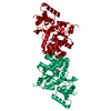 1wq3C 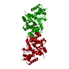 1wq4C  1udf 1udj S: Starting model for refinement C: citing same article ( |
|---|---|
| Similar structure data | |
| Other databases |


-Links
 PDBj
PDBj
- Assembly
| Deposited unit | 
| ||||||||
|---|---|---|---|---|---|---|---|---|---|
| 1 |
| ||||||||
| Unit cell |
|
-Components
| #1: Protein | Mass: 35608.320 Da / Num. of mol.: 2 / Fragment: residues 5-322 / Mutation: Y37V, Q195C Source method: isolated from a genetically manipulated source Source: (gene. exp.) References: UniProt: P00951, UniProt: P0AGJ9*PLUS, tyrosine-tRNA ligase #2: Chemical |   Mass: 509.493 Da / Num. of mol.: 2 / Source method: obtained synthetically / Formula: C19H23N7O8S Mass: 509.493 Da / Num. of mol.: 2 / Source method: obtained synthetically / Formula: C19H23N7O8S#3: Water | ChemComp-HOH / |  Mass: 18.015 Da / Num. of mol.: 115 / Source method: isolated from a natural source / Formula: H2O Mass: 18.015 Da / Num. of mol.: 115 / Source method: isolated from a natural source / Formula: H2O |
|---|
-Experimental details
-Experiment
| Experiment | Method: X-RAY DIFFRACTION / Number of used crystals: 1 |
|---|
- Sample preparation
| Crystal | Density Matthews: 2.54 Å3/Da / Density % sol: 51.21 % |
|---|---|
| Crystal grow | Temperature: 293 K / Method: vapor diffusion, hanging drop / pH: 7.5 Details: ammonium sulphate, PEG 400, sodium chloride, pH 7.5, VAPOR DIFFUSION, HANGING DROP, temperature 293K |
-Data collection
| Diffraction | Mean temperature: 100 K |
|---|---|
| Diffraction source | Source: SYNCHROTRON / Site: SPring-8  / Beamline: BL41XU / Wavelength: 1 Å / Beamline: BL41XU / Wavelength: 1 Å |
| Detector | Type: MARRESEARCH / Detector: CCD / Date: Oct 19, 2003 / Details: mirrors |
| Radiation | Monochromator: Si 111 CHANNEL / Protocol: SINGLE WAVELENGTH / Monochromatic (M) / Laue (L): M / Scattering type: x-ray |
| Radiation wavelength | Wavelength: 1 Å / Relative weight: 1 |
| Reflection | Resolution: 2.7→50 Å / Num. all: 21143 / Num. obs: 20149 / % possible obs: 95.3 % / Observed criterion σ(F): 0 / Observed criterion σ(I): 0 / Redundancy: 6.5 % / Biso Wilson estimate: 12.6 Å2 / Rmerge(I) obs: 0.08 / Rsym value: 0.08 / Net I/σ(I): 15.7 |
| Reflection shell | Resolution: 2.7→2.75 Å / Redundancy: 5.6 % / Rmerge(I) obs: 0.324 / Mean I/σ(I) obs: 2.9 / Num. unique all: 934 / Rsym value: 0.324 / % possible all: 93.4 |
- Processing
| Software |
| ||||||||||||||||||||||||||||||||||||
|---|---|---|---|---|---|---|---|---|---|---|---|---|---|---|---|---|---|---|---|---|---|---|---|---|---|---|---|---|---|---|---|---|---|---|---|---|---|
| Refinement | Method to determine structure: MOLECULAR REPLACEMENT Starting model: PDB ENTRY 1UDF 1udf Resolution: 2.7→46.73 Å / Rfactor Rfree error: 0.006 / Data cutoff high absF: 1849062.93 / Data cutoff low absF: 0 / Isotropic thermal model: RESTRAINED / Cross valid method: THROUGHOUT / σ(F): 0 / σ(I): 0 / Stereochemistry target values: Engh & Huber
| ||||||||||||||||||||||||||||||||||||
| Solvent computation | Solvent model: FLAT MODEL / Bsol: 37.731 Å2 / ksol: 0.31538 e/Å3 | ||||||||||||||||||||||||||||||||||||
| Displacement parameters | Biso mean: 42 Å2
| ||||||||||||||||||||||||||||||||||||
| Refine analyze |
| ||||||||||||||||||||||||||||||||||||
| Refinement step | Cycle: LAST / Resolution: 2.7→46.73 Å
| ||||||||||||||||||||||||||||||||||||
| Refine LS restraints |
| ||||||||||||||||||||||||||||||||||||
| LS refinement shell | Resolution: 2.7→2.87 Å / Rfactor Rfree error: 0.022 / Total num. of bins used: 6
| ||||||||||||||||||||||||||||||||||||
| Xplor file |
|
