 Movie
Movie Controller

[English] 日本語
 Yorodumi
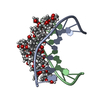
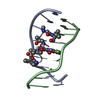
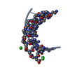
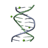
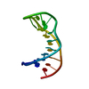
- PDB-1vaq: Crystal structure of the Mg2+-(chromomycin A3)2-d(TTGGCCAA)2 comp... -
+ Open data

- Basic information
| Entry | Database: PDB / ID: 1vaq | ||||||||||||||||||||||||||
|---|---|---|---|---|---|---|---|---|---|---|---|---|---|---|---|---|---|---|---|---|---|---|---|---|---|---|---|
| Title | Crystal structure of the Mg2+-(chromomycin A3)2-d(TTGGCCAA)2 complex reveals GGCC binding specificity of the drug dimer chelated by metal ion | ||||||||||||||||||||||||||
 Components Components | 5'-D(* Keywords KeywordsDNA / Chromomycin A3 / MAD / DNA duplex / GGCC site / DNA kink / CD spectra | Function / homology | Chem-CPH / DNA |  Function and homology information Function and homology informationBiological species | synthetic construct (others) | Method |  X-RAY DIFFRACTION / SYNCHROTRON / MAD / Resolution: 2 Å X-RAY DIFFRACTION / SYNCHROTRON / MAD / Resolution: 2 Å  Authors AuthorsHou, M.H. / Robinson, H. / Gao, Y.G. / Wang, A.H.-J. |  CitationJournal: Nucleic Acids Res. / Year: 2004 CitationJournal: Nucleic Acids Res. / Year: 2004Title: Crystal structure of the [Mg2+-(chromomycin A3)2]-d(TTGGCCAA)2 complex reveals GGCC binding specificity of the drug dimer chelated by a metal ion Authors: Hou, M.H. / Robinson, H. / Gao, Y.G. / Wang, A.H.-J. History |
|
- Structure visualization
| Structure viewer | Molecule: MolmilJmol/JSmol |
|---|
- Downloads & links
-Download
| PDBx/mmCIF format | 1vaq.cif.gz | 45.9 KB | Display | PDBx/mmCIF format |
|---|---|---|---|---|
| PDB format | pdb1vaq.ent.gz | 33.4 KB | Display | PDB format |
| PDBx/mmJSON format | 1vaq.json.gz | Tree view | PDBx/mmJSON format | |
| Others |  Other downloads Other downloads |
-Validation report
| Summary document | 1vaq_validation.pdf.gz | 3.5 MB | Display | wwPDB validaton report |
|---|---|---|---|---|
| Full document | 1vaq_full_validation.pdf.gz | 3.5 MB | Display | |
| Data in XML | 1vaq_validation.xml.gz | 13.5 KB | Display | |
| Data in CIF | 1vaq_validation.cif.gz | 18.3 KB | Display | |
| Arichive directory | https://data.pdbj.org/pub/pdb/validation_reports/va/1vaqftp://data.pdbj.org/pub/pdb/validation_reports/va/1vaq | HTTPS FTP |
-Related structure data
| Similar structure data |
|---|


-Links
 PDBj
PDBj

- Assembly
| Deposited unit | 
| |||||||||||||||
|---|---|---|---|---|---|---|---|---|---|---|---|---|---|---|---|---|
| 1 | 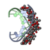
| |||||||||||||||
| 2 | 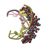
| |||||||||||||||
| Unit cell |
| |||||||||||||||
| Components on special symmetry positions |
|
-Components
-DNA chain , 1 types, 4 molecules ABCD
| #1: DNA chain | Mass: 2426.617 Da / Num. of mol.: 4 / Source method: obtained synthetically Details: The synthetic DNA oligonucleotides were purified by gel electrophoresis. Source: (synth.) synthetic construct (others) |
|---|
-Sugars , 2 types, 8 molecules
| #2: Polysaccharide | 2,6-dideoxy-4-O-methyl-alpha-D-galactopyranose-(1-3)-(2R,3R,6R)-6-hydroxy-2-methyltetrahydro-2H- ...2,6-dideoxy-4-O-methyl-alpha-D-galactopyranose-(1-3)-(2R,3R,6R)-6-hydroxy-2-methyltetrahydro-2H-pyran-3-yl acetate Source method: isolated from a genetically manipulated source #3: Polysaccharide | 3-C-methyl-4-O-acetyl-alpha-L-Olivopyranose-(1-3)-(2R,5S,6R)-6-methyltetrahydro-2H-pyran-2,5-diol- ...3-C-methyl-4-O-acetyl-alpha-L-Olivopyranose-(1-3)-(2R,5S,6R)-6-methyltetrahydro-2H-pyran-2,5-diol-(1-3)-(2R,5S,6R)-6-methyltetrahydro-2H-pyran-2,5-diol Source method: isolated from a genetically manipulated source |
|---|
-Non-polymers , 3 types, 289 molecules 


| #4: Chemical |  Mass: 24.305 Da / Num. of mol.: 2 / Source method: obtained synthetically / Formula: Mg Mass: 24.305 Da / Num. of mol.: 2 / Source method: obtained synthetically / Formula: Mg#5: Chemical | ChemComp-CPH / (  Mass: 420.410 Da / Num. of mol.: 4 / Source method: obtained synthetically / Formula: C21H24O9 Mass: 420.410 Da / Num. of mol.: 4 / Source method: obtained synthetically / Formula: C21H24O9#6: Water | ChemComp-HOH / | Mass: 18.015 Da / Num. of mol.: 283 / Source method: isolated from a natural source / Formula: H2O |
|---|
-Experimental details
-Experiment
| Experiment | Method: X-RAY DIFFRACTION / Number of used crystals: 1 |
|---|
- Sample preparation
| Crystal | Density Matthews: 1.79 Å3/Da / Density % sol: 30.91 % | ||||||||||||||||||||
|---|---|---|---|---|---|---|---|---|---|---|---|---|---|---|---|---|---|---|---|---|---|
| Crystal grow | Temperature: 298 K / Method: vapor diffusion, hanging drop / pH: 5 Details: sodium-cacodylate, MgCl2, pH 5, VAPOR DIFFUSION, HANGING DROP, temperature 298K | ||||||||||||||||||||
| Components of the solutions |
|
-Data collection
| Diffraction | Mean temperature: 110 K |
|---|---|
| Diffraction source | Source: SYNCHROTRON / Site: APS  / Beamline: 19-ID / Wavelength: 1.5432 Å / Beamline: 19-ID / Wavelength: 1.5432 Å |
| Detector | Type: RIGAKU / Detector: IMAGE PLATE |
| Radiation | Monochromator: GRAPHITE / Protocol: MAD / Monochromatic (M) / Laue (L): M / Scattering type: x-ray |
| Radiation wavelength | Wavelength: 1.5432 Å / Relative weight: 1 |
| Reflection | Resolution: 2→50 Å / Num. all: 7815 / Redundancy: 6 % / Rmerge(I) obs: 0.0055 |
- Processing
| Software |
| ||||||||||||||||||||
|---|---|---|---|---|---|---|---|---|---|---|---|---|---|---|---|---|---|---|---|---|---|
| Refinement | Method to determine structure: MAD / Resolution: 2→50 Å / Isotropic thermal model: Isotropic / Cross valid method: THROUGHOUT / σ(F): 10 / Stereochemistry target values: Engh & Huber
| ||||||||||||||||||||
| Refinement step | Cycle: LAST / Resolution: 2→50 Å
| ||||||||||||||||||||
| Refine LS restraints |
| ||||||||||||||||||||
| LS refinement shell | Resolution: 2→2.07 Å /
|
