- PDB-1v57: Crystal Structure of the Disulfide Bond Isomerase DsbG -
+
Open data
ID or keywords:
Loading...
-
Basic information
Entry
Database: PDB / ID: 1v57
Title
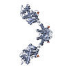
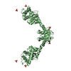
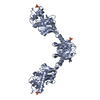
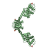
Crystal Structure of the Disulfide Bond Isomerase DsbG
Components
Thiol:disulfide interchange protein dsbG
Keywords
ISOMERASE / oxidized dsbg / redox protein / protein disulfide isomerase / thioredoxin fold / strained redox-active center
Function / homology
Function and homology information
protein disulfide isomerase activity / outer membrane-bounded periplasmic space / protein folding / protein homodimerization activity Similarity search - Function
Single alpha-helices involved in coiled-coils or other helix-helix interfaces - #150 / Disulphide bond isomerase, DsbC/G, N-terminal / Disulphide bond isomerase DsbC/G, N-terminal domain superfamily / Disulphide bond isomerase, DsbC/G, N-terminal / Disulphide bond isomerase, DsbC/G / Disulfide bond isomerase protein N-terminal domain / : / Thioredoxin-like domain / Thioredoxin-like fold / Single alpha-helices involved in coiled-coils or other helix-helix interfaces ...Single alpha-helices involved in coiled-coils or other helix-helix interfaces - #150 / Disulphide bond isomerase, DsbC/G, N-terminal / Disulphide bond isomerase DsbC/G, N-terminal domain superfamily / Disulphide bond isomerase, DsbC/G, N-terminal / Disulphide bond isomerase, DsbC/G / Disulfide bond isomerase protein N-terminal domain / : / Thioredoxin-like domain / Thioredoxin-like fold / Single alpha-helices involved in coiled-coils or other helix-helix interfaces / Nuclear Transport Factor 2; Chain: A, / Helix non-globular / Special / Glutaredoxin / Glutaredoxin / Thioredoxin-like superfamily / Roll / 3-Layer(aba) Sandwich / Alpha Beta Similarity search - Domain/homology
Type: RIGAKU RAXIS IV++ / Detector: IMAGE PLATE / Date: Dec 6, 2001 / Details: OSMIC MAXFLUX CONFOCAL BLUE
Radiation
Monochromator: YALE MIRRORS / Protocol: SINGLE WAVELENGTH / Monochromatic (M) / Laue (L): M / Scattering type: x-ray
Radiation wavelength
Wavelength: 1.5418 Å / Relative weight: 1
Reflection
Resolution: 2→32.17 Å / Num. obs: 36303 / % possible obs: 95.5 % / Observed criterion σ(F): 5 / Redundancy: 3.2 % / Biso Wilson estimate: 22.4 Å2 / Rmerge(I) obs: 0.081 / Net I/σ(I): 6.4
Reflection shell
Resolution: 2→2.07 Å / Redundancy: 2.87 % / Rmerge(I) obs: 0.313 / Mean I/σ(I) obs: 2.1 / Num. unique all: 3323 / % possible all: 88
-
Processing
Software
Name
Version
Classification
CRYSTAL
CLEAR1.3
datacollection
CRYSTAL
CLEAR1.3
datareduction
CNS
refinement
CrystalClear
V. 1.3 (MSC/RIGAKU)
datareduction
CrystalClear
V. 1.3 (MSC/RIGAKU)
datascaling
CNS
phasing
Refinement
Method to determine structure: DIFFERENCE FOURIER Starting model: Structure of Reduced DsbG Resolution: 2→32.17 Å / Isotropic thermal model: isotropic / Cross valid method: THROUGHOUT / σ(F): 0 / Stereochemistry target values: Engh & Huber Details: Refinement: maximum likelihood in CNS. Last minimisation step performed using a reduced weighting for the disulfide bond length.
Rfactor
Num. reflection
% reflection
Selection details
Rfree
0.232
3621
-
RANDOM
Rwork
0.2
-
-
-
all
-
38006
-
-
obs
-
36299
95.4 %
-
Displacement parameters
Biso mean: 28.1 Å2
Baniso -1
Baniso -2
Baniso -3
1-
0.75 Å2
0 Å2
2.72 Å2
2-
-
0.48 Å2
0 Å2
3-
-
-
-1.23 Å2
Refine analyze
Free
Obs
Luzzati coordinate error
0.26 Å
0.22 Å
Luzzati d res low
-
5 Å
Luzzati sigma a
0.29 Å
0.3 Å
Refinement step
Cycle: LAST / Resolution: 2→32.17 Å
Protein
Nucleic acid
Ligand
Solvent
Total
Num. atoms
3799
0
20
509
4328
Refine LS restraints
Refine-ID
Type
Dev ideal
X-RAY DIFFRACTION
c_angle_d
0.005
X-RAY DIFFRACTION
c_angle_deg
1.3
X-RAY DIFFRACTION
c_dihedral_angle_d
22.5
X-RAY DIFFRACTION
c_improper_angle_d
0.84
LS refinement shell
Resolution: 2→2.13 Å / Rfactor Rfree error: 0.014
Rfactor
Num. reflection
% reflection
Rfree
0.315
478
-
Rwork
0.292
-
-
obs
-
4401
77.6 %
+
About Yorodumi
-
News
-
Feb 9, 2022. New format data for meta-information of EMDB entries
New format data for meta-information of EMDB entries
Version 3 of the EMDB header file is now the official format.
The previous official version 1.9 will be removed from the archive.
In the structure databanks used in Yorodumi, some data are registered as the other names, "COVID-19 virus" and "2019-nCoV". Here are the details of the virus and the list of structure data.
Jan 31, 2019. EMDB accession codes are about to change! (news from PDBe EMDB page)
EMDB accession codes are about to change! (news from PDBe EMDB page)
The allocation of 4 digits for EMDB accession codes will soon come to an end. Whilst these codes will remain in use, new EMDB accession codes will include an additional digit and will expand incrementally as the available range of codes is exhausted. The current 4-digit format prefixed with “EMD-” (i.e. EMD-XXXX) will advance to a 5-digit format (i.e. EMD-XXXXX), and so on. It is currently estimated that the 4-digit codes will be depleted around Spring 2019, at which point the 5-digit format will come into force.
The EM Navigator/Yorodumi systems omit the EMD- prefix.
Related info.:Q: What is EMD? / ID/Accession-code notation in Yorodumi/EM Navigator
Yorodumi is a browser for structure data from EMDB, PDB, SASBDB, etc.
This page is also the successor to EM Navigator detail page, and also detail information page/front-end page for Omokage search.
The word "yorodu" (or yorozu) is an old Japanese word meaning "ten thousand". "mi" (miru) is to see.
Related info.:EMDB / PDB / SASBDB / Comparison of 3 databanks / Yorodumi Search / Aug 31, 2016. New EM Navigator & Yorodumi / Yorodumi Papers / Jmol/JSmol / Function and homology information / Changes in new EM Navigator and Yorodumi
 Movie
Movie Controller


 Open data
 Basic information
 Components
 Keywords
 Function and homology information
 X-RAY DIFFRACTION / DIFFERENCE FOURIER / Resolution: 2 Å
  Authors
 Citation
 Structure visualization
 Downloads & links
 Other downloads
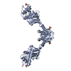


 PDBj
PDBj Assembly
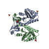

 Mass: 96.063 Da / Num. of mol.: 4 / Source method: obtained synthetically / Formula: SO4
 Mass: 18.015 Da / Num. of mol.: 508 / Source method: isolated from a natural source / Formula: H2O
 Sample preparation
 Processing