 Movie
Movie Controller

[English] 日本語
 Yorodumi
- PDB-1v3p: Crystal structure of d(GCGAGAGC): the DNA octaplex structure with... -
+ Open data

- Basic information
| Entry | Database: PDB / ID: 1v3p | ||||||||||||||||||
|---|---|---|---|---|---|---|---|---|---|---|---|---|---|---|---|---|---|---|---|
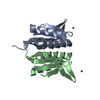
| Title | Crystal structure of d(GCGAGAGC): the DNA octaplex structure with I-motif of G-quartet | ||||||||||||||||||
 Components Components | 5'-D(* Keywords KeywordsDNA / octaplex / quadruplex / G-quartet / I-motif / I-motif of G-quartet / base-intercalated duplex / base-intercalated motif / sheared G:A pair / deoxyribonucleic acid / X-ray analysis | Function / homology | : / DNA |  Function and homology information Function and homology informationMethod |  X-RAY DIFFRACTION / SYNCHROTRON / MOLECULAR REPLACEMENT / Resolution: 2.3 Å X-RAY DIFFRACTION / SYNCHROTRON / MOLECULAR REPLACEMENT / Resolution: 2.3 Å  Authors AuthorsKondo, J. / Umeda, S. / Sunami, T. / Takenaka, A. |  CitationJournal: Nucleic Acids Res. / Year: 2004 CitationJournal: Nucleic Acids Res. / Year: 2004Title: Crystal structures of a DNA octaplex with I-motif of G-quartets and its splitting into two quadruplexes suggest a folding mechanism of eight tandem repeats Authors: Kondo, J. / Adachi, W. / Umeda, S. / Sunami, T. / Takenaka, A. History |
|
- Structure visualization
| Structure viewer | Molecule: MolmilJmol/JSmol |
|---|
- Downloads & links
-Download
| PDBx/mmCIF format | 1v3p.cif.gz | 21.9 KB | Display | PDBx/mmCIF format |
|---|---|---|---|---|
| PDB format | pdb1v3p.ent.gz | 13.8 KB | Display | PDB format |
| PDBx/mmJSON format | 1v3p.json.gz | Tree view | PDBx/mmJSON format | |
| Others |  Other downloads Other downloads |
-Validation report
| Arichive directory | https://data.pdbj.org/pub/pdb/validation_reports/v3/1v3pftp://data.pdbj.org/pub/pdb/validation_reports/v3/1v3p | HTTPS FTP |
|---|
-Related structure data
| Related structure data | 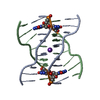 1v3nSC 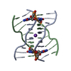 1v3oC S: Starting model for refinement C: citing same article ( |
|---|---|
| Similar structure data |


-Links
 PDBj
PDBj

- Assembly
| Deposited unit | 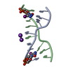
| ||||||||||||||||||
|---|---|---|---|---|---|---|---|---|---|---|---|---|---|---|---|---|---|---|---|
| 1 | 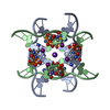
| ||||||||||||||||||
| Unit cell |
| ||||||||||||||||||
| Components on special symmetry positions |
| ||||||||||||||||||
| Details | The biological assembly is an octaplex generated from the duplex in the asymmetric unit by the operations: (x, y, z), (1-x, 1-y, z), (x,1-y,2-z) and (1-x, y, 2-z). |
-Components
| #1: DNA chain | Mass: 2602.540 Da / Num. of mol.: 2 / Source method: obtained synthetically #2: Chemical |   Mass: 39.098 Da / Num. of mol.: 2 / Source method: obtained synthetically / Formula: K Mass: 39.098 Da / Num. of mol.: 2 / Source method: obtained synthetically / Formula: K#3: Water | ChemComp-HOH / |  Mass: 18.015 Da / Num. of mol.: 97 / Source method: isolated from a natural source / Formula: H2O Mass: 18.015 Da / Num. of mol.: 97 / Source method: isolated from a natural source / Formula: H2O |
|---|
-Experimental details
-Experiment
| Experiment | Method: X-RAY DIFFRACTION / Number of used crystals: 1 |
|---|
- Sample preparation
| Crystal | Density Matthews: 1.91 Å3/Da / Density % sol: 35.7 % | ||||||||||||||||||||||||||||||||||||
|---|---|---|---|---|---|---|---|---|---|---|---|---|---|---|---|---|---|---|---|---|---|---|---|---|---|---|---|---|---|---|---|---|---|---|---|---|---|
| Crystal grow | Temperature: 277 K / Method: vapor diffusion, hanging drop / pH: 7 Details: 2-methyl-2,4-pentanediol, spermine tetrahydrochloride, sodium chloride, potassium chloride, sodium cacodylate , pH 7.0, VAPOR DIFFUSION, HANGING DROP, temperature 277K | ||||||||||||||||||||||||||||||||||||
| Components of the solutions |
|
-Data collection
| Diffraction | Mean temperature: 100 K |
|---|---|
| Diffraction source | Source: SYNCHROTRON / Site: Photon Factory  / Beamline: BL-18B / Wavelength: 1 Å / Beamline: BL-18B / Wavelength: 1 Å |
| Detector | Type: ADSC QUANTUM 4 / Detector: CCD / Date: Nov 11, 2000 |
| Radiation | Protocol: SINGLE WAVELENGTH / Monochromatic (M) / Laue (L): M / Scattering type: x-ray |
| Radiation wavelength | Wavelength: 1 Å / Relative weight: 1 |
| Reflection | Resolution: 2.3→20 Å / Num. all: 2014 / Num. obs: 1980 / % possible obs: 98.4 % / Observed criterion σ(F): 0 / Observed criterion σ(I): 0 / Rmerge(I) obs: 0.084 / Net I/σ(I): 7.1 |
| Reflection shell | Resolution: 2.3→2.38 Å / Rmerge(I) obs: 0.24 / Mean I/σ(I) obs: 2.4 / Num. unique all: 199 / % possible all: 99 |
- Processing
| Software |
| |||||||||||||||||||||
|---|---|---|---|---|---|---|---|---|---|---|---|---|---|---|---|---|---|---|---|---|---|---|
| Refinement | Method to determine structure: MOLECULAR REPLACEMENT Starting model: PDB ENTRY 1V3N Resolution: 2.3→9 Å / σ(F): 3 Stereochemistry target values: G. PARKINSON ET AL., (1996) ACTACRYST. D52, 57-64
| |||||||||||||||||||||
| Refine analyze | Luzzati coordinate error obs: 0.31 Å / Luzzati d res low obs: 5 Å / Luzzati sigma a obs: 0.29 Å | |||||||||||||||||||||
| Refinement step | Cycle: LAST / Resolution: 2.3→9 Å
| |||||||||||||||||||||
| Refine LS restraints |
| |||||||||||||||||||||
| LS refinement shell | Resolution: 2.3→2.38 Å
|
