 Movie
Movie Controller

[English] 日本語
 Yorodumi
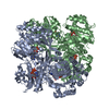
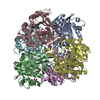
- PDB-1v1a: 2-KETO-3-DEOXYGLUCONATE KINASE FROM THERMUS THERMOPHILUS WITH BOU... -
+ Open data

- Basic information
| Entry | Database: PDB / ID: 1v1a | ||||||
|---|---|---|---|---|---|---|---|
| Title | 2-KETO-3-DEOXYGLUCONATE KINASE FROM THERMUS THERMOPHILUS WITH BOUND 2-KETO-3-DEOXYGLUCONATE AND ADP | ||||||
 Components Components | 2-KETO-3-DEOXYGLUCONATE KINASE | ||||||
 Keywords Keywords | TRANSFERASE / 2-KETO-3-DEOXYGLUCONATE KINASE / ATP / THERMUS THERMOPHILUS / STRUCTURAL GENOMICS / RIKEN STRUCTURAL GENOMICS/PROTEOMICS INITIATIVE / RSGI | ||||||
| Function / homology |  Function and homology information Function and homology information2-dehydro-3-deoxygluconokinase / 2-dehydro-3-deoxygluconokinase activity / phosphorylation / nucleotide binding / ATP binding Similarity search - Function | ||||||
| Biological species |   THERMUS THERMOPHILUS (bacteria) THERMUS THERMOPHILUS (bacteria) | ||||||
| Method |  X-RAY DIFFRACTION / SYNCHROTRON / MOLECULAR REPLACEMENT / Resolution: 2.1 Å X-RAY DIFFRACTION / SYNCHROTRON / MOLECULAR REPLACEMENT / Resolution: 2.1 Å | ||||||
 Authors Authors | Tahirov, T.H. / Inagaki, E. | ||||||
 Citation Citation | Journal: J. Mol. Biol. / Year: 2004 Title: Structure of Thermus thermophilus 2-Keto-3-deoxygluconate kinase: evidence for recognition of an open chain substrate. Authors: Ohshima, N. / Inagaki, E. / Yasuike, K. / Takio, K. / Tahirov, T.H. #1: Journal: Acta Crystallogr.,Sect.D / Year: 2004 Title: Crystallization and Preliminary Crystallographic Analysis of 2-Keto-3-Deoxygluconate Kinase from Thermus Thermophilus. Authors: Inagaki, E. / Ukita, Y. / Kumei, M. / Kajihara, Y. / Tahirov, T.H. | ||||||
| History |
|
- Structure visualization
| Structure viewer | Molecule: MolmilJmol/JSmol |
|---|
- Downloads & links
-Download
| PDBx/mmCIF format | 1v1a.cif.gz | 132 KB | Display | PDBx/mmCIF format |
|---|---|---|---|---|
| PDB format | pdb1v1a.ent.gz | 104.1 KB | Display | PDB format |
| PDBx/mmJSON format | 1v1a.json.gz | Tree view | PDBx/mmJSON format | |
| Others |  Other downloads Other downloads |
-Validation report
| Arichive directory | https://data.pdbj.org/pub/pdb/validation_reports/v1/1v1aftp://data.pdbj.org/pub/pdb/validation_reports/v1/1v1a | HTTPS FTP |
|---|
-Related structure data


-Links
 PDBj
PDBj- Assembly
| Deposited unit | 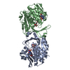
| ||||||||
|---|---|---|---|---|---|---|---|---|---|
| 1 | 
| ||||||||
| Unit cell |
|
-Components
| #1: Protein | Mass: 33470.184 Da / Num. of mol.: 2 Source method: isolated from a genetically manipulated source Source: (gene. exp.) THERMUS THERMOPHILUS (bacteria) / Strain: HB8 / Plasmid: PET11A / Production host: #2: Chemical |   Mass: 178.140 Da / Num. of mol.: 2 / Source method: obtained synthetically / Formula: C6H10O6 Mass: 178.140 Da / Num. of mol.: 2 / Source method: obtained synthetically / Formula: C6H10O6#3: Chemical |   Mass: 427.201 Da / Num. of mol.: 2 / Source method: obtained synthetically / Formula: C10H15N5O10P2 / Comment: ADP, energy-carrying molecule*YM Mass: 427.201 Da / Num. of mol.: 2 / Source method: obtained synthetically / Formula: C10H15N5O10P2 / Comment: ADP, energy-carrying molecule*YM#4: Water | ChemComp-HOH / |  Mass: 18.015 Da / Num. of mol.: 211 / Source method: isolated from a natural source / Formula: H2O Mass: 18.015 Da / Num. of mol.: 211 / Source method: isolated from a natural source / Formula: H2O |
|---|
-Experimental details
-Experiment
| Experiment | Method: X-RAY DIFFRACTION / Number of used crystals: 1 |
|---|
- Sample preparation
| Crystal | Density Matthews: 2.53 Å3/Da / Density % sol: 52.5 % |
|---|---|
| Crystal grow | Temperature: 298 K / Method: vapor diffusion, sitting drop / pH: 8.5 Details: VAPOUR-DIFFUSION SITTING DROP AT 298 K. 10.0 MG/ML OF PROTEIN SOLUTION CONTAINING 5 MM KDG AND 5 MM AMP-PNP WAS MIXED WITH RESERVOIR SOLUTION CONTAINING 0.35 M AMMONIUM SULFATE AND 0.1 M ...Details: VAPOUR-DIFFUSION SITTING DROP AT 298 K. 10.0 MG/ML OF PROTEIN SOLUTION CONTAINING 5 MM KDG AND 5 MM AMP-PNP WAS MIXED WITH RESERVOIR SOLUTION CONTAINING 0.35 M AMMONIUM SULFATE AND 0.1 M TRIS-HCL BUFFER, PH 8.5. BEFORE THE DATA COLLECTION THE CRYSTAL WAS SOAKED IN SOLUTION CONTAINING 0.35 M MAGNESIUM CHLORIDE INSTEAD OF AMMONIUM SULFATE. THE CRYOPROTECTANT CONTAINED 33% OF ETHYLENE GLYCOL IN ADDITION TO MAGNESIUM CHLORIDE, BUFFER AND LIGANDS |
-Data collection
| Diffraction | Mean temperature: 100 K |
|---|---|
| Diffraction source | Source: SYNCHROTRON / Site: SPring-8  / Beamline: BL26B1 / Wavelength: 1 / Beamline: BL26B1 / Wavelength: 1 |
| Detector | Type: RIGAKU IMAGE PLATE R-AXISV / Detector: IMAGE PLATE / Date: Mar 15, 2003 |
| Radiation | Protocol: SINGLE WAVELENGTH / Monochromatic (M) / Laue (L): M / Scattering type: x-ray |
| Radiation wavelength | Wavelength: 1 Å / Relative weight: 1 |
| Reflection | Resolution: 2.1→30 Å / Num. obs: 38730 / % possible obs: 97.9 % / Observed criterion σ(I): -0.4 / Redundancy: 4.58 % / Biso Wilson estimate: 13.1 Å2 / Rmerge(I) obs: 0.056 / Net I/σ(I): 19.6 |
| Reflection shell | Resolution: 2.1→2.18 Å / Rmerge(I) obs: 0.355 / Mean I/σ(I) obs: 2.68 / % possible all: 96.2 |
- Processing
| Software |
| ||||||||||||||||||||||||||||||||||||||||||||||||||||||||||||||||||||||||||||||||
|---|---|---|---|---|---|---|---|---|---|---|---|---|---|---|---|---|---|---|---|---|---|---|---|---|---|---|---|---|---|---|---|---|---|---|---|---|---|---|---|---|---|---|---|---|---|---|---|---|---|---|---|---|---|---|---|---|---|---|---|---|---|---|---|---|---|---|---|---|---|---|---|---|---|---|---|---|---|---|---|---|---|
| Refinement | Method to determine structure: MOLECULAR REPLACEMENT Starting model: PDB ENTRY V19 Resolution: 2.1→29.82 Å / Rfactor Rfree error: 0.008 / Data cutoff high absF: 1289180.4 / Isotropic thermal model: RESTRAINED / Cross valid method: THROUGHOUT / σ(F): 0 Details: CRYSTAL IS WITH HEMIHEDRAL TWINNING, TWINNING OPERATOR IS "H, -H-K, -L", TWINNING FRACTION IS 0.5
| ||||||||||||||||||||||||||||||||||||||||||||||||||||||||||||||||||||||||||||||||
| Solvent computation | Solvent model: FLAT MODEL / Bsol: 85.7706 Å2 / ksol: 0.355934 e/Å3 | ||||||||||||||||||||||||||||||||||||||||||||||||||||||||||||||||||||||||||||||||
| Displacement parameters | Biso mean: 51.4 Å2
| ||||||||||||||||||||||||||||||||||||||||||||||||||||||||||||||||||||||||||||||||
| Refine analyze |
| ||||||||||||||||||||||||||||||||||||||||||||||||||||||||||||||||||||||||||||||||
| Refinement step | Cycle: LAST / Resolution: 2.1→29.82 Å
| ||||||||||||||||||||||||||||||||||||||||||||||||||||||||||||||||||||||||||||||||
| Refine LS restraints |
| ||||||||||||||||||||||||||||||||||||||||||||||||||||||||||||||||||||||||||||||||
| LS refinement shell | Resolution: 2.1→2.18 Å / Rfactor Rfree error: 0.023 / Total num. of bins used: 10
| ||||||||||||||||||||||||||||||||||||||||||||||||||||||||||||||||||||||||||||||||
| Xplor file |
|
