 Movie
Movie Controller

[English] 日本語
 Yorodumi
- PDB-1ux1: Bacillus subtilis cytidine deaminase with a Cys53His and an Arg56... -
+ Open data

- Basic information
| Entry | Database: PDB / ID: 1ux1 | ||||||
|---|---|---|---|---|---|---|---|
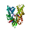
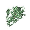
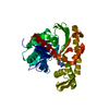
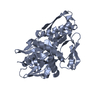
| Title | Bacillus subtilis cytidine deaminase with a Cys53His and an Arg56Gln substitution | ||||||
 Components Components | CYTIDINE DEAMINASE | ||||||
 Keywords Keywords | HYDROLASE / CYTIDINE DEAMINASE / CDD / TETRAMER / ZINC BINDING / PYRIMIDINE METABOLISM / SALVAGE | ||||||
| Function / homology |  Function and homology information Function and homology informationpyrimidine-containing compound metabolic process / cytidine deaminase / nucleobase-containing small molecule metabolic process / cytidine deaminase activity / zinc ion binding / identical protein binding / cytosol Similarity search - Function | ||||||
| Biological species |  | ||||||
| Method |  X-RAY DIFFRACTION / MOLECULAR REPLACEMENT / Resolution: 2.36 Å X-RAY DIFFRACTION / MOLECULAR REPLACEMENT / Resolution: 2.36 Å | ||||||
 Authors Authors | Johansson, E. / Neuhard, J. / Willemoes, M. / Larsen, S. | ||||||
 Citation Citation | Journal: Biochemistry / Year: 2004 Title: Structural, Kinetic, and Mutational Studies of the Zinc Ion Environment in Tetrameric Cytidine Deaminase Authors: Johansson, E. / Neuhard, J. / Willemoes, M. / Larsen, S. | ||||||
| History |
|
- Structure visualization
| Structure viewer | Molecule: MolmilJmol/JSmol |
|---|
- Downloads & links
-Download
| PDBx/mmCIF format | 1ux1.cif.gz | 115.1 KB | Display | PDBx/mmCIF format |
|---|---|---|---|---|
| PDB format | pdb1ux1.ent.gz | 88.6 KB | Display | PDB format |
| PDBx/mmJSON format | 1ux1.json.gz | Tree view | PDBx/mmJSON format | |
| Others |  Other downloads Other downloads |
-Validation report
| Arichive directory | https://data.pdbj.org/pub/pdb/validation_reports/ux/1ux1ftp://data.pdbj.org/pub/pdb/validation_reports/ux/1ux1 | HTTPS FTP |
|---|
-Related structure data
| Related structure data |  1uwzC 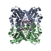 1ux0C 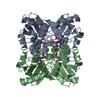 1jtkS S: Starting model for refinement C: citing same article ( |
|---|---|
| Similar structure data |


-Links
 PDBj
PDBj- Assembly
| Deposited unit | 
| ||||||||||||||||
|---|---|---|---|---|---|---|---|---|---|---|---|---|---|---|---|---|---|
| 1 |
| ||||||||||||||||
| Unit cell |
| ||||||||||||||||
| Components on special symmetry positions |
| ||||||||||||||||
| Noncrystallographic symmetry (NCS) | NCS oper:
|
-Components
| #1: Protein | Mass: 14874.952 Da / Num. of mol.: 4 / Mutation: YES Source method: isolated from a genetically manipulated source Source: (gene. exp.) #2: Chemical | ChemComp-THU /   Mass: 230.218 Da / Num. of mol.: 4 / Source method: obtained synthetically / Formula: C9H14N2O5 Mass: 230.218 Da / Num. of mol.: 4 / Source method: obtained synthetically / Formula: C9H14N2O5#3: Chemical | ChemComp-ZN /   Mass: 65.409 Da / Num. of mol.: 4 / Source method: obtained synthetically / Formula: Zn Mass: 65.409 Da / Num. of mol.: 4 / Source method: obtained synthetically / Formula: Zn#4: Chemical | ChemComp-TRS / |   Mass: 122.143 Da / Num. of mol.: 1 / Source method: obtained synthetically / Formula: C4H12NO3 / Comment: pH buffer*YM Mass: 122.143 Da / Num. of mol.: 1 / Source method: obtained synthetically / Formula: C4H12NO3 / Comment: pH buffer*YM#5: Water | ChemComp-HOH / |  Mass: 18.015 Da / Num. of mol.: 98 / Source method: isolated from a natural source / Formula: H2O Mass: 18.015 Da / Num. of mol.: 98 / Source method: isolated from a natural source / Formula: H2OCompound details | ENGINEERED MUTATION ARG 56 GLN IN CHAINS A B C D ENGINEERED MUTATION CYS 53 HIS IN CHAINS A B C D ...ENGINEERED | |
|---|
-Experimental details
-Experiment
| Experiment | Method: X-RAY DIFFRACTION / Number of used crystals: 1 |
|---|
- Sample preparation
| Crystal | Density Matthews: 2.1 Å3/Da / Density % sol: 41.4 % |
|---|---|
| Crystal grow | Method: vapor diffusion / pH: 7.5 Details: VAPOUR DIFFUSION AT RT: PROTEIN: 4.6 MG/ML, + 5 MM TETRAHYDRODEOXYURIDINE, PRECIPITANT: 30 % PEG400, 0.2M MAGNESIUM CHLORIDE, 0.1 M HEPES PH 7.5 |
-Data collection
| Diffraction | Mean temperature: 120 K |
|---|---|
| Diffraction source | Source: ROTATING ANODE / Type: RIGAKU / Wavelength: 1.542 |
| Detector | Type: MARRESEARCH / Detector: IMAGE PLATE / Date: Sep 23, 2002 / Details: OSMIC MIRRORS |
| Radiation | Protocol: SINGLE WAVELENGTH / Monochromatic (M) / Laue (L): M / Scattering type: x-ray |
| Radiation wavelength | Wavelength: 1.542 Å / Relative weight: 1 |
| Reflection | Resolution: 2.36→25 Å / Num. obs: 21401 / % possible obs: 98.9 % / Redundancy: 6.8 % / Biso Wilson estimate: 28.3 Å2 / Rmerge(I) obs: 0.059 / Net I/σ(I): 29.8 |
| Reflection shell | Resolution: 2.36→2.41 Å / Rmerge(I) obs: 0.217 / Mean I/σ(I) obs: 6.9 / % possible all: 83.9 |
- Processing
| Software |
| ||||||||||||||||||||||||||||||||||||||||||||||||||||||||||||||||||||||||||||||||
|---|---|---|---|---|---|---|---|---|---|---|---|---|---|---|---|---|---|---|---|---|---|---|---|---|---|---|---|---|---|---|---|---|---|---|---|---|---|---|---|---|---|---|---|---|---|---|---|---|---|---|---|---|---|---|---|---|---|---|---|---|---|---|---|---|---|---|---|---|---|---|---|---|---|---|---|---|---|---|---|---|---|
| Refinement | Method to determine structure: MOLECULAR REPLACEMENT Starting model: PDB ENTRY 1JTK Resolution: 2.36→24.66 Å / Rfactor Rfree error: 0.007 / Data cutoff high absF: 2004857.7 / Isotropic thermal model: RESTRAINED / Cross valid method: THROUGHOUT / σ(F): 0
| ||||||||||||||||||||||||||||||||||||||||||||||||||||||||||||||||||||||||||||||||
| Solvent computation | Solvent model: FLAT MODEL / Bsol: 30.6894 Å2 / ksol: 0.346096 e/Å3 | ||||||||||||||||||||||||||||||||||||||||||||||||||||||||||||||||||||||||||||||||
| Displacement parameters | Biso mean: 32.6 Å2
| ||||||||||||||||||||||||||||||||||||||||||||||||||||||||||||||||||||||||||||||||
| Refine analyze |
| ||||||||||||||||||||||||||||||||||||||||||||||||||||||||||||||||||||||||||||||||
| Refinement step | Cycle: LAST / Resolution: 2.36→24.66 Å
| ||||||||||||||||||||||||||||||||||||||||||||||||||||||||||||||||||||||||||||||||
| Refine LS restraints |
| ||||||||||||||||||||||||||||||||||||||||||||||||||||||||||||||||||||||||||||||||
| LS refinement shell | Resolution: 2.36→2.51 Å / Rfactor Rfree error: 0.022 / Total num. of bins used: 6
| ||||||||||||||||||||||||||||||||||||||||||||||||||||||||||||||||||||||||||||||||
| Xplor file |
|
