 Movie
Movie Controller


+ Open data

- Basic information
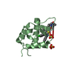
| Entry | Database: PDB / ID: 1urr | ||||||
|---|---|---|---|---|---|---|---|
| Title | A novel Drosophila Melanogaster Acylphosphatase (AcPDro2) | ||||||
 Components Components | CG18505 PROTEIN | ||||||
 Keywords Keywords | HYDROLASE / ACYLPHOSPHATASE / ENZYME | ||||||
| Function / homology |  Function and homology information Function and homology informationacylphosphatase / acylphosphatase activity / hydrolase activity / cytoplasm Similarity search - Function | ||||||
| Biological species |  | ||||||
| Method |  X-RAY DIFFRACTION / SYNCHROTRON / MOLECULAR REPLACEMENT / Resolution: 1.5 Å X-RAY DIFFRACTION / SYNCHROTRON / MOLECULAR REPLACEMENT / Resolution: 1.5 Å | ||||||
 Authors Authors | Rosano, C. / Zuccotti, S. / Bolognesi, M. | ||||||
 Citation Citation | Journal: Acta Crystallogr.,Sect.D / Year: 2004 Title: Three-Dimensional Structural Characterization of a Novel Drosophila Melanogaster Acylphosphatase Authors: Zuccotti, S. / Rosano, C. / Ramazzotti, M. / Degl'Innocenti, D. / Stefani, M. / Manao, G. / Bolognesi, M. #1: Journal: FEBS Lett. / Year: 2003 Title: Characterization of a Novel Drosophila Melanogaster Acylphosphatase Authors: Degl'Innocenti, D. / Ramazzotti, M. / Marzocchini, R. / Chiti, F. / Raugei, G. | ||||||
| History |
| ||||||
| Remark 700 | SHEET THE SHEET STRUCTURE OF THIS MOLECULE IS BIFURCATED. IN ORDER TO REPRESENT THIS FEATURE IN ... SHEET THE SHEET STRUCTURE OF THIS MOLECULE IS BIFURCATED. IN ORDER TO REPRESENT THIS FEATURE IN THE SHEET RECORDS BELOW, TWO SHEETS ARE DEFINED. |
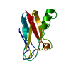
- Structure visualization
| Structure viewer | Molecule: MolmilJmol/JSmol |
|---|
- Downloads & links
-Download
| PDBx/mmCIF format | 1urr.cif.gz | 58.7 KB | Display | PDBx/mmCIF format |
|---|---|---|---|---|
| PDB format | pdb1urr.ent.gz | 42.7 KB | Display | PDB format |
| PDBx/mmJSON format | 1urr.json.gz | Tree view | PDBx/mmJSON format | |
| Others |  Other downloads Other downloads |
-Validation report
| Arichive directory | https://data.pdbj.org/pub/pdb/validation_reports/ur/1urrftp://data.pdbj.org/pub/pdb/validation_reports/ur/1urr | HTTPS FTP |
|---|
-Related structure data
| Related structure data |  2acyS S: Starting model for refinement |
|---|---|
| Similar structure data |


-Links
 PDBj
PDBj- Assembly
| Deposited unit | 
| ||||||||
|---|---|---|---|---|---|---|---|---|---|
| 1 |
| ||||||||
| Unit cell |
| ||||||||
| Components on special symmetry positions |
|
-Components
| #1: Protein | Mass: 11783.540 Da / Num. of mol.: 1 Source method: isolated from a genetically manipulated source Source: (gene. exp.)  |
|---|---|
| #2: Chemical | ChemComp-GOL /   Mass: 92.094 Da / Num. of mol.: 1 / Source method: obtained synthetically / Formula: C3H8O3 Mass: 92.094 Da / Num. of mol.: 1 / Source method: obtained synthetically / Formula: C3H8O3 |
| #3: Water | ChemComp-HOH /  Mass: 18.015 Da / Num. of mol.: 109 / Source method: isolated from a natural source / Formula: H2O Mass: 18.015 Da / Num. of mol.: 109 / Source method: isolated from a natural source / Formula: H2O |
-Experimental details
-Experiment
| Experiment | Method: X-RAY DIFFRACTION / Number of used crystals: 1 |
|---|
- Sample preparation
| Crystal | Density Matthews: 2.52 Å3/Da / Density % sol: 55.2 % |
|---|---|
| Crystal grow | pH: 4.5 / Details: pH 4.50 |
-Data collection
| Diffraction | Mean temperature: 100 K |
|---|---|
| Diffraction source | Source: SYNCHROTRON / Site: ESRF  / Beamline: ID29 / Wavelength: 0.933 / Beamline: ID29 / Wavelength: 0.933 |
| Radiation | Protocol: SINGLE WAVELENGTH / Monochromatic (M) / Laue (L): M / Scattering type: x-ray |
| Radiation wavelength | Wavelength: 0.933 Å / Relative weight: 1 |
| Reflection | Resolution: 1.5→39.5 Å / Num. obs: 19859 / % possible obs: 98.7 % / Observed criterion σ(I): 3 / Redundancy: 6.2 % / Biso Wilson estimate: 29.5 Å2 / Rmerge(I) obs: 0.047 / Net I/σ(I): 17.2 |
| Reflection shell | Resolution: 1.5→1.55 Å / Rmerge(I) obs: 0.3 / Mean I/σ(I) obs: 2.9 / % possible all: 97.1 |
- Processing
| Software |
| ||||||||||||||||||||
|---|---|---|---|---|---|---|---|---|---|---|---|---|---|---|---|---|---|---|---|---|---|
| Refinement | Method to determine structure: MOLECULAR REPLACEMENT Starting model: PDB ENTRY 2ACY Resolution: 1.5→10 Å / SU ML: 0.047 / ESU R Free: 0.079
| ||||||||||||||||||||
| Displacement parameters | Biso mean: 26.9 Å2 | ||||||||||||||||||||
| Refinement step | Cycle: LAST / Resolution: 1.5→10 Å
|
