 Movie
Movie Controller

[English] 日本語
 Yorodumi
- PDB-1uhm: Solution structure of the globular domain of linker histone homol... -
+ Open data

- Basic information
| Entry | Database: PDB / ID: 1uhm | ||||||
|---|---|---|---|---|---|---|---|
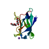
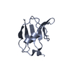
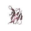
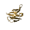
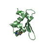
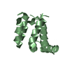
| Title | Solution structure of the globular domain of linker histone homolog Hho1p from S. cerevisiae | ||||||
 Components Components | Histone H1 | ||||||
 Keywords Keywords | STRUCTURAL PROTEIN / winged helix-turn-helix / linker histone / S. cerevisiae / RIKEN Structural Genomics/Proteomics Initiative / RSGI / Structural Genomics | ||||||
| Function / homology |  Function and homology information Function and homology informationsporulation / negative regulation of DNA recombination / regulation of double-strand break repair / chromosome condensation / supercoiled DNA binding / nucleosomal DNA binding / phosphate ion binding / protein-DNA complex / chromatin DNA binding / structural constituent of chromatin ...sporulation / negative regulation of DNA recombination / regulation of double-strand break repair / chromosome condensation / supercoiled DNA binding / nucleosomal DNA binding / phosphate ion binding / protein-DNA complex / chromatin DNA binding / structural constituent of chromatin / nucleosome / nucleosome assembly / double-stranded DNA binding / molecular adaptor activity / nucleic acid binding / regulation of DNA-templated transcription / chromatin / DNA binding / nucleus Similarity search - Function | ||||||
| Biological species |  | ||||||
| Method | SOLUTION NMR / distance geometry simulated annealing, molecular dynamics | ||||||
 Authors Authors | Ono, K. / Kusano, O. / Shimotakahara, S. / Shimizu, M. / Yamazaki, T. / Shindo, H. / RIKEN Structural Genomics/Proteomics Initiative (RSGI) | ||||||
 Citation Citation | Journal: Nucleic Acids Res. / Year: 2003 Title: The linker histone homolog Hho1p from Saccharomyces cerevisiae represents a winged helix-turn-helix fold as determined by NMR spectroscopy. Authors: Ono, K. / Kusano, O. / Shimotakahara, S. / Shimizu, M. / Yamazaki, T. / Shindo, H. | ||||||
| History |
|
- Structure visualization
| Structure viewer | Molecule: MolmilJmol/JSmol |
|---|
- Downloads & links
-Download
| PDBx/mmCIF format | 1uhm.cif.gz | 475.2 KB | Display | PDBx/mmCIF format |
|---|---|---|---|---|
| PDB format | pdb1uhm.ent.gz | 396.7 KB | Display | PDB format |
| PDBx/mmJSON format | 1uhm.json.gz | Tree view | PDBx/mmJSON format | |
| Others |  Other downloads Other downloads |
-Validation report
| Arichive directory | https://data.pdbj.org/pub/pdb/validation_reports/uh/1uhmftp://data.pdbj.org/pub/pdb/validation_reports/uh/1uhm | HTTPS FTP |
|---|
-Related structure data
| Similar structure data | |
|---|---|
| Other databases |


-Links
 PDBj
PDBj- Assembly
| Deposited unit | 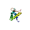
| |||||||||
|---|---|---|---|---|---|---|---|---|---|---|
| 1 |
| |||||||||
| NMR ensembles |
|
-Components
| #1: Protein | Mass: 8516.781 Da / Num. of mol.: 1 / Fragment: globular domain Source method: isolated from a genetically manipulated source Source: (gene. exp.) Gene: HHO1 / Plasmid: pET21b / Species (production host): Escherichia coli / Production host:  |
|---|
-Experimental details
-Experiment
| Experiment | Method: SOLUTION NMR |
|---|
- Sample preparation
| Details | Contents: 1mM HD1 U-15N,13C; 10mM phosphate buffer / Solvent system: 90% H2O/10% D2O |
|---|---|
| Sample conditions | pH: 6.2 / Pressure: ambient / Temperature: 298 K |
| Crystal grow | *PLUS Method: other / Details: NMR |
-NMR measurement
| Radiation | Protocol: SINGLE WAVELENGTH / Monochromatic (M) / Laue (L): M |
|---|---|
| Radiation wavelength | Relative weight: 1 |
| NMR spectrometer | Type: Bruker DMX / Manufacturer: Bruker / Model: DMX / Field strength: 750 MHz |
- Processing
| NMR software | Name:  X-PLOR / Version: 3.1 / Developer: Brunger, A.T. / Classification: refinement X-PLOR / Version: 3.1 / Developer: Brunger, A.T. / Classification: refinement |
|---|---|
| Refinement | Method: distance geometry simulated annealing, molecular dynamics Software ordinal: 1 |
| NMR ensemble | Conformer selection criteria: structures with acceptable covalent geometry, structures with favorable non-bond energy, structures with the least restraint violations, structures with the lowest energy Conformers calculated total number: 100 / Conformers submitted total number: 20 |
