 Movie
Movie Controller

[English] 日本語
 Yorodumi
- PDB-1ue6: Crystal structure of the single-stranded dna-binding protein from... -
+ Open data

- Basic information
| Entry | Database: PDB / ID: 1ue6 | ||||||
|---|---|---|---|---|---|---|---|
| Title | Crystal structure of the single-stranded dna-binding protein from mycobacterium tuberculosis | ||||||
 Components Components | Single-strand binding protein | ||||||
 Keywords Keywords | DNA BINDING PROTEIN / OLIGONUCLEOTIDE BINDING FOLD / DNA-BINDING PROTEIN / Structural Genomics / PSI / Protein Structure Initiative / TB Structural Genomics Consortium / TBSGC | ||||||
| Function / homology |  Function and homology information Function and homology informationnucleoid / enzyme activator activity / single-stranded DNA binding / DNA replication / response to antibiotic / DNA damage response / extracellular region / plasma membrane Similarity search - Function | ||||||
| Biological species |   Mycobacterium tuberculosis (bacteria) Mycobacterium tuberculosis (bacteria) | ||||||
| Method |  X-RAY DIFFRACTION / MOLECULAR REPLACEMENT / Resolution: 2.7 Å X-RAY DIFFRACTION / MOLECULAR REPLACEMENT / Resolution: 2.7 Å | ||||||
 Authors Authors | Saikrishnan, K. / Jeyakanthan, J. / Venkatesh, J. / Acharya, N. / Sekar, K. / Varshney, U. / Vijayan, M. / TB Structural Genomics Consortium (TBSGC) | ||||||
 Citation Citation | Journal: J.MOL.BIOL. / Year: 2003 Title: Structure of Mycobacterium tuberculosis single-stranded DNA-binding protein. Variability in quaternary structure and its implications Authors: Saikrishnan, K. / Jeyakanthan, J. / Venkatesh, J. / Acharya, N. / Sekar, K. / Varshney, U. / Vijayan, M. #1: Journal: ACTA CRYSTALLOGR.,SECT.D / Year: 2002 Title: Crystallization and preliminary X-ray studies of the single-stranded DNA-binding protein from Mycobacterium tuberculosis. Authors: Saikrishnan, K. / Jeyakanthan, J. / Venkatesh, J. / Acharya, N. / Purnapatre, K. / Sekar, K. / Varshney, U. / Vijayan, M. | ||||||
| History |
|
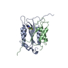
- Structure visualization
| Structure viewer | Molecule: MolmilJmol/JSmol |
|---|
- Downloads & links
-Download
| PDBx/mmCIF format | 1ue6.cif.gz | 98 KB | Display | PDBx/mmCIF format |
|---|---|---|---|---|
| PDB format | pdb1ue6.ent.gz | 74.3 KB | Display | PDB format |
| PDBx/mmJSON format | 1ue6.json.gz | Tree view | PDBx/mmJSON format | |
| Others |  Other downloads Other downloads |
-Validation report
| Arichive directory | https://data.pdbj.org/pub/pdb/validation_reports/ue/1ue6ftp://data.pdbj.org/pub/pdb/validation_reports/ue/1ue6 | HTTPS FTP |
|---|
-Related structure data
| Related structure data |  1ue1SC  1ue5C  1ue7C S: Starting model for refinement C: citing same article ( |
|---|---|
| Similar structure data | |
| Other databases |
-Links
 PDBj
PDBj- Assembly
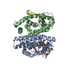
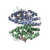
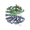
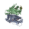
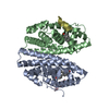
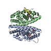
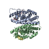
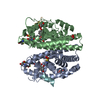
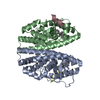
| Deposited unit | 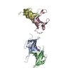
| ||||||||
|---|---|---|---|---|---|---|---|---|---|
| 1 | 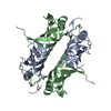
| ||||||||
| 2 | 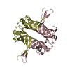
| ||||||||
| Unit cell |
|
-Components
| #1: Protein | Mass: 17372.066 Da / Num. of mol.: 4 Source method: isolated from a genetically manipulated source Source: (gene. exp.) Mycobacterium tuberculosis (bacteria) / Plasmid: PET11d / Production host: #2: Water | ChemComp-HOH / |  Mass: 18.015 Da / Num. of mol.: 229 / Source method: isolated from a natural source / Formula: H2O Mass: 18.015 Da / Num. of mol.: 229 / Source method: isolated from a natural source / Formula: H2O |
|---|
-Experimental details
-Experiment
| Experiment | Method: X-RAY DIFFRACTION / Number of used crystals: 1 |
|---|
- Sample preparation
| Crystal | Density Matthews: 3.38 Å3/Da / Density % sol: 63.3 % | ||||||||||||||||||||||||||||||||||||||||||||||||||||||||
|---|---|---|---|---|---|---|---|---|---|---|---|---|---|---|---|---|---|---|---|---|---|---|---|---|---|---|---|---|---|---|---|---|---|---|---|---|---|---|---|---|---|---|---|---|---|---|---|---|---|---|---|---|---|---|---|---|---|
| Crystal grow | Temperature: 298 K / Method: vapor diffusion, hanging drop / pH: 7.4 Details: 2M MAGNESIUM CHLORIDE, 500mM SODIUM CHLORIDE, 20mM TRIS-HCL, pH 7.4, VAPOR DIFFUSION, HANGING DROP, temperature 298K | ||||||||||||||||||||||||||||||||||||||||||||||||||||||||
| Crystal grow | *PLUS Temperature: 298 K / Method: vapor diffusion, hanging drop / Details: Saikrishnan, K., (2002) Acta Cryst., D58, 327. | ||||||||||||||||||||||||||||||||||||||||||||||||||||||||
| Components of the solutions | *PLUS
|
-Data collection
| Diffraction | Mean temperature: 100 K |
|---|---|
| Diffraction source | Source: ROTATING ANODE / Type: RIGAKU RU200 / Wavelength: 1.5418 Å |
| Detector | Type: MARRESEARCH / Detector: IMAGE PLATE / Date: Jul 30, 2001 |
| Radiation | Protocol: SINGLE WAVELENGTH / Monochromatic (M) / Laue (L): M / Scattering type: x-ray |
| Radiation wavelength | Wavelength: 1.5418 Å / Relative weight: 1 |
| Reflection | Resolution: 2.7→20 Å / Num. all: 75505 / Num. obs: 17174 / % possible obs: 97.9 % / Observed criterion σ(F): 1 / Observed criterion σ(I): 1 / Biso Wilson estimate: 42.5 Å2 / Rmerge(I) obs: 0.073 |
| Reflection shell | Resolution: 2.7→2.8 Å / % possible all: 97.6 |
| Reflection | *PLUS Highest resolution: 2.7 Å / Redundancy: 4.4 % |
| Reflection shell | *PLUS % possible obs: 97.6 % / Redundancy: 4.4 % / Rmerge(I) obs: 0.582 |
- Processing
| Software |
| ||||||||||||||||||||||||||||||||||||
|---|---|---|---|---|---|---|---|---|---|---|---|---|---|---|---|---|---|---|---|---|---|---|---|---|---|---|---|---|---|---|---|---|---|---|---|---|---|
| Refinement | Method to determine structure: MOLECULAR REPLACEMENT Starting model: 1UE1 Resolution: 2.7→15 Å / Rfactor Rfree error: 0.01 / Data cutoff high absF: 205124.33 / Data cutoff low absF: 0 / Isotropic thermal model: RESTRAINED / Cross valid method: THROUGHOUT / σ(F): 0 / Stereochemistry target values: Engh & Huber Details: A LARGE NUMBER OF THE ISOLATED WATER MOLECULES REPRESENT THE DISCREET AND ISOLATED ELECTRON DENSITIES, WHICH MAY CORRESPOND TO THE UNDEFINED REGIONS OF THE POLYPEPTIDE CHAIN PRIMARILY AT THE ...Details: A LARGE NUMBER OF THE ISOLATED WATER MOLECULES REPRESENT THE DISCREET AND ISOLATED ELECTRON DENSITIES, WHICH MAY CORRESPOND TO THE UNDEFINED REGIONS OF THE POLYPEPTIDE CHAIN PRIMARILY AT THE C-TERMINUS AND THE LOOPS. The crystals were obtained from a sample containing truncated protein. the nature of the truncation is not known.
| ||||||||||||||||||||||||||||||||||||
| Solvent computation | Solvent model: FLAT MODEL / Bsol: 59.224 Å2 / ksol: 0.290218 e/Å3 | ||||||||||||||||||||||||||||||||||||
| Displacement parameters | Biso mean: 57.6 Å2
| ||||||||||||||||||||||||||||||||||||
| Refine analyze |
| ||||||||||||||||||||||||||||||||||||
| Refinement step | Cycle: LAST / Resolution: 2.7→15 Å
| ||||||||||||||||||||||||||||||||||||
| Refine LS restraints |
| ||||||||||||||||||||||||||||||||||||
| LS refinement shell | Resolution: 2.7→2.87 Å / Rfactor Rfree error: 0.034 / Total num. of bins used: 6
| ||||||||||||||||||||||||||||||||||||
| Xplor file |
| ||||||||||||||||||||||||||||||||||||
| Refinement | *PLUS Lowest resolution: 15 Å | ||||||||||||||||||||||||||||||||||||
| Solvent computation | *PLUS | ||||||||||||||||||||||||||||||||||||
| Displacement parameters | *PLUS | ||||||||||||||||||||||||||||||||||||
| Refine LS restraints | *PLUS
|
