- PDB-1u8x: CRYSTAL STRUCTURE OF GLVA FROM BACILLUS SUBTILIS, A METAL-REQUIRI... -
+
Open data
ID or keywords:
Loading...
-
Basic information
Entry
Database: PDB / ID: 1u8x
Title
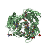
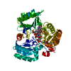
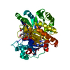
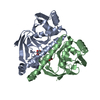
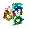
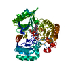
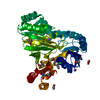
CRYSTAL STRUCTURE OF GLVA FROM BACILLUS SUBTILIS, A METAL-REQUIRING, NAD-DEPENDENT 6-PHOSPHO-ALPHA-GLUCOSIDASE
Components
Maltose-6'-phosphate glucosidase
Keywords
HYDROLASE / STRUCTURAL GENOMICS / PSI / protein structure initiative / MCSG / GLUCOSIDASE / NAD-DEPENDENT / Midwest Center for Structural Genomics
Function / homology
Function and homology information
maltose-6'-phosphate glucosidase / maltose-6'-phosphate glucosidase activity / oxidoreductase activity, acting on the CH-OH group of donors, NAD or NADP as acceptor / hydrolase activity, hydrolyzing O-glycosyl compounds / carbohydrate metabolic process / metal ion binding / cytosol Similarity search - Function
Glycoside hydrolase, family 4, conserved site / Glycosyl hydrolases family 4 signature. / Glycoside hydrolase, family 4 / Glycosyl hydrolase, family 4, C-terminal / Family 4 glycosyl hydrolase / Family 4 glycosyl hydrolase C-terminal domain / L-2-Hydroxyisocaproate Dehydrogenase; Chain A, domain 2 / Lactate dehydrogenase/glycoside hydrolase, family 4, C-terminal / Lactate dehydrogenase/glycoside hydrolase, family 4, C-terminal / NAD(P)-binding Rossmann-like Domain ...Glycoside hydrolase, family 4, conserved site / Glycosyl hydrolases family 4 signature. / Glycoside hydrolase, family 4 / Glycosyl hydrolase, family 4, C-terminal / Family 4 glycosyl hydrolase / Family 4 glycosyl hydrolase C-terminal domain / L-2-Hydroxyisocaproate Dehydrogenase; Chain A, domain 2 / Lactate dehydrogenase/glycoside hydrolase, family 4, C-terminal / Lactate dehydrogenase/glycoside hydrolase, family 4, C-terminal / NAD(P)-binding Rossmann-like Domain / NAD(P)-binding domain superfamily / Alpha-Beta Complex / Rossmann fold / 3-Layer(aba) Sandwich / Alpha Beta Similarity search - Domain/homology
In the structure databanks used in Yorodumi, some data are registered as the other names, "COVID-19 virus" and "2019-nCoV". Here are the details of the virus and the list of structure data.
Jan 31, 2019. EMDB accession codes are about to change! (news from PDBe EMDB page)
EMDB accession codes are about to change! (news from PDBe EMDB page)
The allocation of 4 digits for EMDB accession codes will soon come to an end. Whilst these codes will remain in use, new EMDB accession codes will include an additional digit and will expand incrementally as the available range of codes is exhausted. The current 4-digit format prefixed with “EMD-” (i.e. EMD-XXXX) will advance to a 5-digit format (i.e. EMD-XXXXX), and so on. It is currently estimated that the 4-digit codes will be depleted around Spring 2019, at which point the 5-digit format will come into force.
The EM Navigator/Yorodumi systems omit the EMD- prefix.
Related info.:Q: What is EMD? / ID/Accession-code notation in Yorodumi/EM Navigator
Yorodumi is a browser for structure data from EMDB, PDB, SASBDB, etc.
This page is also the successor to EM Navigator detail page, and also detail information page/front-end page for Omokage search.
The word "yorodu" (or yorozu) is an old Japanese word meaning "ten thousand". "mi" (miru) is to see.
Related info.:EMDB / PDB / SASBDB / Comparison of 3 databanks / Yorodumi Search / Aug 31, 2016. New EM Navigator & Yorodumi / Yorodumi Papers / Jmol/JSmol / Function and homology information / Changes in new EM Navigator and Yorodumi
 Movie
Movie Controller

 Yorodumi
 Open data
 Basic information
 Components
 Keywords
 Function and homology information
 X-RAY DIFFRACTION /
  Authors
 Citation
 Structure visualization
 Downloads & links
 Other downloads
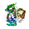

 PDBj
PDBj


 Assembly
 Type: D-saccharide, alpha linking / Mass: 260.136 Da / Num. of mol.: 1
 Mass: 54.938 Da / Num. of mol.: 1 / Source method: obtained synthetically / Formula: Mn
 Mass: 663.425 Da / Num. of mol.: 1 / Source method: obtained synthetically / Formula: C21H27N7O14P2 / Comment: NAD*YM
 Mass: 18.015 Da / Num. of mol.: 162 / Source method: isolated from a natural source / Formula: H2O
 Sample preparation
 / Beamline: 5ID-B / Wavelength: 0.979 Å
 Processing