 Movie
Movie Controller

[English] 日本語
 Yorodumi
- PDB-1u6i: The Structure of native coenzyme F420-dependent methylenetetrahyd... -
+ Open data

- Basic information
| Entry | Database: PDB / ID: 1u6i | ||||||
|---|---|---|---|---|---|---|---|
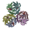
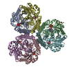
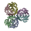
| Title | The Structure of native coenzyme F420-dependent methylenetetrahydromethanopterin dehydrogenase at 2.2A resolution | ||||||
 Components Components | F420-dependent methylenetetrahydromethanopterin dehydrogenase | ||||||
 Keywords Keywords | OXIDOREDUCTASE / MONOMER: ALPHA/BETA DOMAIN / HELIX BUNDLE / TRIMER OF DIMERS | ||||||
| Function / homology |  Function and homology information Function and homology informationmethylenetetrahydromethanopterin dehydrogenase / methylenetetrahydromethanopterin dehydrogenase activity / methanogenesis, from carbon dioxide / ferredoxin hydrogenase activity / one-carbon metabolic process Similarity search - Function | ||||||
| Biological species |   Methanopyrus kandleri (archaea) Methanopyrus kandleri (archaea) | ||||||
| Method |  X-RAY DIFFRACTION / SYNCHROTRON / Resolution: 2.2 Å X-RAY DIFFRACTION / SYNCHROTRON / Resolution: 2.2 Å | ||||||
 Authors Authors | Warkentin, E. / Hagemeier, C.H. / Shima, S. / Thauer, R.K. / Ermler, U. | ||||||
 Citation Citation | Journal: Acta Crystallogr.,Sect.D / Year: 2005 Title: The structure of F420-dependent methylenetetrahydromethanopterin dehydrogenase: a crystallographic 'superstructure' of the selenomethionine-labelled protein crystal structure. Authors: Warkentin, E. / Hagemeier, C.H. / Shima, S. / Thauer, R.K. / Ermler, U. #1: Journal: J.Mol.Biol. / Year: 2003Title: Coenzyme F420-dependent methylenetetrahydromethanopterin dehydrogenase (Mtd) from Methanopyrus kandleri: a methanogenic enzyme with an unusual quarternary structure. Authors: Hagemeier, C.H. / Shima, S. / Thauer, R.K. / Bourenkov, G. / Bartunik, H.D. / Ermler, U. | ||||||
| History |
|
- Structure visualization
| Structure viewer | Molecule: MolmilJmol/JSmol |
|---|
- Downloads & links
-Download
| PDBx/mmCIF format | 1u6i.cif.gz | 643 KB | Display | PDBx/mmCIF format |
|---|---|---|---|---|
| PDB format | pdb1u6i.ent.gz | 535.2 KB | Display | PDB format |
| PDBx/mmJSON format | 1u6i.json.gz | Tree view | PDBx/mmJSON format | |
| Others |  Other downloads Other downloads |
-Validation report
| Arichive directory | https://data.pdbj.org/pub/pdb/validation_reports/u6/1u6iftp://data.pdbj.org/pub/pdb/validation_reports/u6/1u6i | HTTPS FTP |
|---|
-Related structure data
| Related structure data |  1u6jC  1u6kC 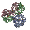 1qv9S S: Starting model for refinement C: citing same article ( |
|---|---|
| Similar structure data |


-Links
 PDBj
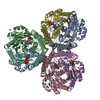
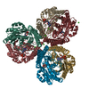
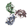
PDBj- Assembly
| Deposited unit | 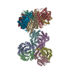
| ||||||||
|---|---|---|---|---|---|---|---|---|---|
| 1 | 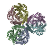
| ||||||||
| 2 | 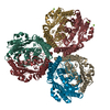
| ||||||||
| Unit cell |
|
-Components
| #1: Protein | Mass: 31415.885 Da / Num. of mol.: 12 Source method: isolated from a genetically manipulated source Source: (gene. exp.) Methanopyrus kandleri (archaea) / Gene: mtd / Plasmid: pET17b / Species (production host): Escherichia coli / Production host:  #2: Chemical | ChemComp-MG /   Mass: 24.305 Da / Num. of mol.: 8 / Source method: obtained synthetically / Formula: Mg Mass: 24.305 Da / Num. of mol.: 8 / Source method: obtained synthetically / Formula: Mg#3: Water | ChemComp-HOH / |  Mass: 18.015 Da / Num. of mol.: 666 / Source method: isolated from a natural source / Formula: H2O Mass: 18.015 Da / Num. of mol.: 666 / Source method: isolated from a natural source / Formula: H2O |
|---|
-Experimental details
-Experiment
| Experiment | Method: X-RAY DIFFRACTION / Number of used crystals: 1 |
|---|
- Sample preparation
| Crystal | Density Matthews: 2.6 Å3/Da / Density % sol: 48.4 % |
|---|---|
| Crystal grow | Temperature: 277 K / Method: vapor diffusion, hanging drop / pH: 7 Details: MOPS, KOH, MPD, PEG 400, Mg acetate, Na phosphate, pH 7.0, VAPOR DIFFUSION, HANGING DROP, temperature 277K |
-Data collection
| Diffraction | Mean temperature: 100 K |
|---|---|
| Diffraction source | Source: SYNCHROTRON / Site: ESRF  / Beamline: ID14-4 / Wavelength: 0.9393 Å / Beamline: ID14-4 / Wavelength: 0.9393 Å |
| Detector | Type: ADSC QUANTUM 4 / Detector: CCD / Date: Jul 11, 2002 |
| Radiation | Monochromator: DIAMOND / Protocol: SINGLE WAVELENGTH / Monochromatic (M) / Laue (L): M / Scattering type: x-ray |
| Radiation wavelength | Wavelength: 0.9393 Å / Relative weight: 1 |
| Reflection | Resolution: 2.2→20 Å / Num. obs: 190905 / % possible obs: 96.7 % / Observed criterion σ(F): 0 / Observed criterion σ(I): -1 / Redundancy: 1.86 % / Biso Wilson estimate: 17.6 Å2 / Rmerge(I) obs: 0.141 / Rsym value: 0.064 / Net I/σ(I): 6.5 |
| Reflection shell | Resolution: 2.2→2.4 Å / Redundancy: 1.8 % / Rmerge(I) obs: 0.312 / Mean I/σ(I) obs: 4.4 / Num. unique all: 38875 / Rsym value: 0.129 / % possible all: 93.6 |
- Processing
| Software |
| ||||||||||||||||||||
|---|---|---|---|---|---|---|---|---|---|---|---|---|---|---|---|---|---|---|---|---|---|
| Refinement | Starting model: Selenomethionine-labelled methylenetetrahydromethanopterin dehydrogenase (MTD), PDB-ID 1QV9 Resolution: 2.2→19.35 Å / Rfactor Rfree error: 0.002 / Data cutoff high absF: 9714980.33 / Data cutoff low absF: 0 / Isotropic thermal model: RESTRAINED / Cross valid method: THROUGHOUT / σ(F): 0 / Stereochemistry target values: Engh & Huber Details: Description of the kMtd(Se) structure (PDB-ID 1QV9; space group C2221) in C2, a, subgroup of C2221
| ||||||||||||||||||||
| Solvent computation | Solvent model: FLAT MODEL / Bsol: 46.3 Å2 / ksol: 0.352 e/Å3 | ||||||||||||||||||||
| Displacement parameters | Biso mean: 39.1 Å2
| ||||||||||||||||||||
| Refine analyze |
| ||||||||||||||||||||
| Refinement step | Cycle: LAST / Resolution: 2.2→19.35 Å
| ||||||||||||||||||||
| Refine LS restraints |
| ||||||||||||||||||||
| Refine LS restraints NCS | NCS model details: THE MAIN-CHAIN ATOMS OF THE 2ND HEXAMER WERE STRONGLY TIED TO THE FIRST, THE SIDE-CHAIN ATOMS LESS TIGHT. IN ADDITION THE (PSEUDO) TWO-FOLD ALONG THE A AXIS WAS WEAKLY IMPOSED. | ||||||||||||||||||||
| LS refinement shell | Resolution: 2.2→2.37 Å / Rfactor Rfree error: 0.008 / Total num. of bins used: 6
| ||||||||||||||||||||
| Xplor file |
|
