 Movie
Movie Controller


+ Open data

- Basic information
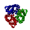
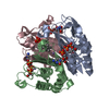
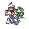
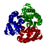
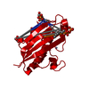
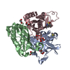
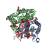
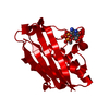
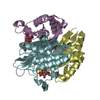
| Entry | Database: PDB / ID: 1u3l | ||||||
|---|---|---|---|---|---|---|---|
| Title | IspF with Mg and CDP | ||||||
 Components Components | 2-C-methyl-D-erythritol 2,4-cyclodiphosphate synthase | ||||||
 Keywords Keywords | LYASE / MEP pathway / terpene biosynthesis | ||||||
| Function / homology |  Function and homology information Function and homology information2-C-methyl-D-erythritol 2,4-cyclodiphosphate synthase / 2-C-methyl-D-erythritol 2,4-cyclodiphosphate synthase activity / isopentenyl diphosphate biosynthetic process, methylerythritol 4-phosphate pathway / terpenoid biosynthetic process / ubiquinone biosynthetic process / manganese ion binding / zinc ion binding / metal ion binding / identical protein binding Similarity search - Function | ||||||
| Biological species |  | ||||||
| Method |  X-RAY DIFFRACTION / FOURIER SYNTHESIS / Resolution: 2.5 Å X-RAY DIFFRACTION / FOURIER SYNTHESIS / Resolution: 2.5 Å | ||||||
 Authors Authors | Steinbacher, S. / Kaiser, J. / Wungsintaweekul, J. / Hechst, S. / Eisenreich, W. / Gerhardt, S. / Bacher, A. / Rohdich, F. | ||||||
 Citation Citation | Journal: J.Mol.Biol. / Year: 2002 Title: Structure of 2C-Methyl-D-Erythritol-2,4-Cyclodiphosphate Synthase Involved in Mevalonate Independent Biosynthesis of Isoprenoids Authors: Steinbacher, S. / Kaiser, J. / Wungsintaweekul, J. / Hechst, S. / Eisenreich, W. / Gerhardt, S. / Bacher, A. / Rohdich, F. | ||||||
| History |
|
- Structure visualization
| Structure viewer | Molecule: MolmilJmol/JSmol |
|---|
- Downloads & links
-Download
| PDBx/mmCIF format | 1u3l.cif.gz | 43.4 KB | Display | PDBx/mmCIF format |
|---|---|---|---|---|
| PDB format | pdb1u3l.ent.gz | 29.8 KB | Display | PDB format |
| PDBx/mmJSON format | 1u3l.json.gz | Tree view | PDBx/mmJSON format | |
| Others |  Other downloads Other downloads |
-Validation report
| Arichive directory | https://data.pdbj.org/pub/pdb/validation_reports/u3/1u3lftp://data.pdbj.org/pub/pdb/validation_reports/u3/1u3l | HTTPS FTP |
|---|
-Related structure data
| Related structure data |  1jy8SC  1u3pC  1u40C  1u43C S: Starting model for refinement C: citing same article ( |
|---|---|
| Similar structure data |

-Links
 PDBj
PDBj- Assembly
| Deposited unit | 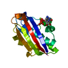
| ||||||||
|---|---|---|---|---|---|---|---|---|---|
| 1 | 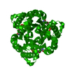
| ||||||||
| Unit cell |
| ||||||||
| Details | Trimer generated by crystallographic three-fold x,y,z y,z,x z,x,y |
-Components
| #1: Protein | Mass: 16920.531 Da / Num. of mol.: 1 Source method: isolated from a genetically manipulated source Source: (gene. exp.) References: UniProt: P62617, 2-C-methyl-D-erythritol 2,4-cyclodiphosphate synthase |
|---|---|
| #2: Chemical | ChemComp-MG /   Mass: 24.305 Da / Num. of mol.: 1 / Source method: obtained synthetically / Formula: Mg Mass: 24.305 Da / Num. of mol.: 1 / Source method: obtained synthetically / Formula: Mg |
| #3: Chemical | ChemComp-ZN /   Mass: 65.409 Da / Num. of mol.: 1 / Source method: obtained synthetically / Formula: Zn Mass: 65.409 Da / Num. of mol.: 1 / Source method: obtained synthetically / Formula: Zn |
| #4: Chemical | ChemComp-CDP / 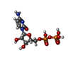  Mass: 403.176 Da / Num. of mol.: 1 / Source method: obtained synthetically / Formula: C9H15N3O11P2 Mass: 403.176 Da / Num. of mol.: 1 / Source method: obtained synthetically / Formula: C9H15N3O11P2 |
-Experimental details
-Experiment
| Experiment | Method: X-RAY DIFFRACTION / Number of used crystals: 1 |
|---|
- Sample preparation
| Crystal | Density Matthews: 7.45 Å3/Da / Density % sol: 84 % |
|---|---|
| Crystal grow | Temperature: 293 K / Method: vapor diffusion, sitting drop / pH: 7 Details: ammonium formiate, pH 7, VAPOR DIFFUSION, SITTING DROP, temperature 293K |
-Data collection
| Diffraction | Mean temperature: 100 K |
|---|---|
| Diffraction source | Source: ROTATING ANODE / Type: RIGAKU RU200 / Wavelength: 1.541 Å |
| Detector | Type: MARRESEARCH / Detector: IMAGE PLATE / Date: Mar 1, 2001 |
| Radiation | Protocol: SINGLE WAVELENGTH / Monochromatic (M) / Laue (L): M / Scattering type: x-ray |
| Radiation wavelength | Wavelength: 1.541 Å / Relative weight: 1 |
| Reflection | Resolution: 2.5→25 Å / Num. all: 17015 / Num. obs: 17015 / % possible obs: 97.3 % / Observed criterion σ(F): 0 / Observed criterion σ(I): 0 / Redundancy: 3.2 % / Rmerge(I) obs: 0.064 |
- Processing
| Software |
| |||||||||||||||||||||
|---|---|---|---|---|---|---|---|---|---|---|---|---|---|---|---|---|---|---|---|---|---|---|
| Refinement | Method to determine structure: FOURIER SYNTHESIS Starting model: 1JY8 Resolution: 2.5→8 Å / Cross valid method: THROUGHOUT / σ(F): 0 / Stereochemistry target values: Engh & Huber
| |||||||||||||||||||||
| Refinement step | Cycle: LAST / Resolution: 2.5→8 Å
| |||||||||||||||||||||
| Refine LS restraints |
|
