 Movie
Movie Controller

[English] 日本語
 Yorodumi
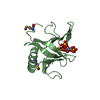
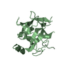
- PDB-1u2b: Triglycine variant of the Grp1 Pleckstrin Homology Domain unliganded -
+ Open data

- Basic information
| Entry | Database: PDB / ID: 1u2b | ||||||
|---|---|---|---|---|---|---|---|
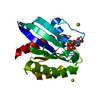
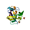
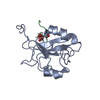
| Title | Triglycine variant of the Grp1 Pleckstrin Homology Domain unliganded | ||||||
 Components Components | Cytohesin 3 | ||||||
 Keywords Keywords | LIPID BINDING PROTEIN / PH domain / lipid binding / phosphoinositides | ||||||
| Function / homology |  Function and homology information Function and homology informationIntra-Golgi traffic / establishment of epithelial cell polarity / Golgi vesicle transport / regulation of ARF protein signal transduction / phosphatidylinositol-3,4,5-trisphosphate binding / bicellular tight junction / ruffle / positive regulation of cell adhesion / guanyl-nucleotide exchange factor activity / adherens junction ...Intra-Golgi traffic / establishment of epithelial cell polarity / Golgi vesicle transport / regulation of ARF protein signal transduction / phosphatidylinositol-3,4,5-trisphosphate binding / bicellular tight junction / ruffle / positive regulation of cell adhesion / guanyl-nucleotide exchange factor activity / adherens junction / nucleoplasm / plasma membrane / cytoplasm / cytosol Similarity search - Function | ||||||
| Biological species |  | ||||||
| Method |  X-RAY DIFFRACTION / MOLECULAR REPLACEMENT / Resolution: 1.8 Å X-RAY DIFFRACTION / MOLECULAR REPLACEMENT / Resolution: 1.8 Å | ||||||
 Authors Authors | Cronin, T.C. / DiNitto, J.P. / Czech, M.P. / Lambright, D.G. | ||||||
 Citation Citation | Journal: Embo J. / Year: 2004 Title: Structural determinants of phosphoinositide selectivity in splice variants of Grp1 family PH domains Authors: Cronin, T.C. / DiNitto, J.P. / Czech, M.P. / Lambright, D.G. | ||||||
| History |
|
- Structure visualization
| Structure viewer | Molecule: MolmilJmol/JSmol |
|---|
- Downloads & links
-Download
| PDBx/mmCIF format | 1u2b.cif.gz | 42.5 KB | Display | PDBx/mmCIF format |
|---|---|---|---|---|
| PDB format | pdb1u2b.ent.gz | 29 KB | Display | PDB format |
| PDBx/mmJSON format | 1u2b.json.gz | Tree view | PDBx/mmJSON format | |
| Others |  Other downloads Other downloads |
-Validation report
| Arichive directory | https://data.pdbj.org/pub/pdb/validation_reports/u2/1u2bftp://data.pdbj.org/pub/pdb/validation_reports/u2/1u2b | HTTPS FTP |
|---|
-Related structure data
| Related structure data |  1u27C 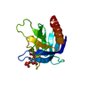 1u29C 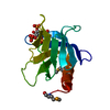 1fgyS C: citing same article ( S: Starting model for refinement |
|---|---|
| Similar structure data |


-Links
 PDBj
PDBj
- Assembly
| Deposited unit | 
| ||||||||
|---|---|---|---|---|---|---|---|---|---|
| 1 |
| ||||||||
| Unit cell |
|
-Components
| #1: Protein | Mass: 16259.445 Da / Num. of mol.: 1 / Fragment: PH Source method: isolated from a genetically manipulated source Source: (gene. exp.)  | ||
|---|---|---|---|
| #2: Chemical |   Mass: 96.063 Da / Num. of mol.: 2 / Source method: obtained synthetically / Formula: SO4 Mass: 96.063 Da / Num. of mol.: 2 / Source method: obtained synthetically / Formula: SO4#3: Water | ChemComp-HOH / |  Mass: 18.015 Da / Num. of mol.: 76 / Source method: isolated from a natural source / Formula: H2O Mass: 18.015 Da / Num. of mol.: 76 / Source method: isolated from a natural source / Formula: H2O |
-Experimental details
-Experiment
| Experiment | Method: X-RAY DIFFRACTION / Number of used crystals: 1 |
|---|
- Sample preparation
| Crystal | Density Matthews: 2.73 Å3/Da / Density % sol: 55 % |
|---|---|
| Crystal grow | Temperature: 293 K / Method: vapor diffusion, hanging drop / pH: 4.2 Details: PEG 4000, sodium acetate, 10% glycerol, lithium sulfate, pH 4.2, VAPOR DIFFUSION, HANGING DROP, temperature 293K |
-Data collection
| Diffraction | Mean temperature: 100 K |
|---|---|
| Diffraction source | Source: ROTATING ANODE / Wavelength: 1.54 Å |
| Detector | Type: MARRESEARCH / Detector: IMAGE PLATE / Date: Sep 19, 2000 |
| Radiation | Protocol: SINGLE WAVELENGTH / Monochromatic (M) / Laue (L): M / Scattering type: x-ray |
| Radiation wavelength | Wavelength: 1.54 Å / Relative weight: 1 |
| Reflection | Resolution: 1.8→50 Å / Num. all: 15937 / Num. obs: 15719 / % possible obs: 98.6 % / Observed criterion σ(F): 0 / Observed criterion σ(I): -3 |
| Reflection shell | Resolution: 1.8→1.85 Å / % possible all: 96.8 |
- Processing
| Software |
| ||||||||||||||||||||||||||||||||||||||||||||||||||||||||||||||||||||||||||||||||||||||||||||||||||||
|---|---|---|---|---|---|---|---|---|---|---|---|---|---|---|---|---|---|---|---|---|---|---|---|---|---|---|---|---|---|---|---|---|---|---|---|---|---|---|---|---|---|---|---|---|---|---|---|---|---|---|---|---|---|---|---|---|---|---|---|---|---|---|---|---|---|---|---|---|---|---|---|---|---|---|---|---|---|---|---|---|---|---|---|---|---|---|---|---|---|---|---|---|---|---|---|---|---|---|---|---|---|
| Refinement | Method to determine structure: MOLECULAR REPLACEMENT Starting model: 1FGY Resolution: 1.8→20 Å / Cor.coef. Fo:Fc: 0.939 / Cor.coef. Fo:Fc free: 0.924 / SU B: 2.746 / SU ML: 0.086 / Cross valid method: THROUGHOUT / σ(F): 0 / σ(I): 0 / ESU R: 0.128 / ESU R Free: 0.119 / Stereochemistry target values: MAXIMUM LIKELIHOOD Details: Hydrogens have been added in the riding positions. Several atoms for residues 280, 323, 334, 335, 336, 353, 355, 370 and 387 were modeled into this structure with zero occupancy because they ...Details: Hydrogens have been added in the riding positions. Several atoms for residues 280, 323, 334, 335, 336, 353, 355, 370 and 387 were modeled into this structure with zero occupancy because they are part of the PH domain and were not deleted or mutated.
| ||||||||||||||||||||||||||||||||||||||||||||||||||||||||||||||||||||||||||||||||||||||||||||||||||||
| Solvent computation | Ion probe radii: 0.8 Å / Shrinkage radii: 0.8 Å / VDW probe radii: 1.4 Å / Solvent model: BABINET MODEL WITH MASK | ||||||||||||||||||||||||||||||||||||||||||||||||||||||||||||||||||||||||||||||||||||||||||||||||||||
| Displacement parameters | Biso mean: 23.028 Å2
| ||||||||||||||||||||||||||||||||||||||||||||||||||||||||||||||||||||||||||||||||||||||||||||||||||||
| Refinement step | Cycle: LAST / Resolution: 1.8→20 Å
| ||||||||||||||||||||||||||||||||||||||||||||||||||||||||||||||||||||||||||||||||||||||||||||||||||||
| Refine LS restraints |
| ||||||||||||||||||||||||||||||||||||||||||||||||||||||||||||||||||||||||||||||||||||||||||||||||||||
| LS refinement shell | Resolution: 1.8→1.846 Å / Total num. of bins used: 20 /
|
