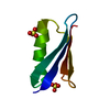
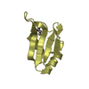
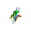
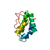
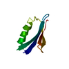
- PDB-1tp4: Solution structure of the XPC binding domain of hHR23A protein -
+
Open data
ID or keywords:
Loading...
-
Basic information
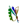
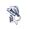
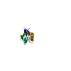
Entry
Database: PDB / ID: 1tp4
Title
Solution structure of the XPC binding domain of hHR23A protein
Components
UV excision repair protein RAD23 homolog A
Keywords
DNA REPAIR / NER / XPC / Rad23
Function / homology
Function and homology information
regulation of proteasomal ubiquitin-dependent protein catabolic process / histone H4K20 demethylase activity / Oxidoreductases; Acting on paired donors, with incorporation or reduction of molecular oxygen; With 2-oxoglutarate as one donor, and incorporation of one atom of oxygen into each donor / ubiquitin-specific protease binding / proteasome binding / polyubiquitin modification-dependent protein binding / positive regulation of viral genome replication / positive regulation of cell cycle / proteasome complex / Josephin domain DUBs ...regulation of proteasomal ubiquitin-dependent protein catabolic process / histone H4K20 demethylase activity / Oxidoreductases; Acting on paired donors, with incorporation or reduction of molecular oxygen; With 2-oxoglutarate as one donor, and incorporation of one atom of oxygen into each donor / ubiquitin-specific protease binding / proteasome binding / polyubiquitin modification-dependent protein binding / positive regulation of viral genome replication / positive regulation of cell cycle / proteasome complex / Josephin domain DUBs / ubiquitin binding / nucleotide-excision repair / protein destabilization / DNA Damage Recognition in GG-NER / kinase binding / Formation of Incision Complex in GG-NER / positive regulation of proteasomal ubiquitin-dependent protein catabolic process / single-stranded DNA binding / damaged DNA binding / proteasome-mediated ubiquitin-dependent protein catabolic process / protein-containing complex / nucleoplasm / nucleus / cytosol / cytoplasm Similarity search - Function
Protocol: SINGLE WAVELENGTH / Monochromatic (M) / Laue (L): M
Radiation wavelength
Relative weight: 1
NMR spectrometer
Type: Bruker DRX / Manufacturer: Bruker / Model: DRX / Field strength: 600 MHz
-
Processing
NMR software
Name
Version
Classification
XwinNMR
2.6
collection
Felix
2000
dataanalysis
X-PLOR
3.1
structuresolution
X-PLOR
3.1
refinement
Refinement
Method: distance geometry / Software ordinal: 1 Details: The structures are based on a total of 917 non-redundant NOE-derived distance constraints, 82 dihedral angle restraints and 42 distance restraints from hydrogen bonds.
NMR representative
Selection criteria: lowest energy
NMR ensemble
Conformer selection criteria: structures with the lowest energy Conformers calculated total number: 100 / Conformers submitted total number: 25
+
About Yorodumi
-
News
-
Feb 9, 2022. New format data for meta-information of EMDB entries
New format data for meta-information of EMDB entries
Version 3 of the EMDB header file is now the official format.
The previous official version 1.9 will be removed from the archive.
In the structure databanks used in Yorodumi, some data are registered as the other names, "COVID-19 virus" and "2019-nCoV". Here are the details of the virus and the list of structure data.
Jan 31, 2019. EMDB accession codes are about to change! (news from PDBe EMDB page)
EMDB accession codes are about to change! (news from PDBe EMDB page)
The allocation of 4 digits for EMDB accession codes will soon come to an end. Whilst these codes will remain in use, new EMDB accession codes will include an additional digit and will expand incrementally as the available range of codes is exhausted. The current 4-digit format prefixed with “EMD-” (i.e. EMD-XXXX) will advance to a 5-digit format (i.e. EMD-XXXXX), and so on. It is currently estimated that the 4-digit codes will be depleted around Spring 2019, at which point the 5-digit format will come into force.
The EM Navigator/Yorodumi systems omit the EMD- prefix.
Related info.:Q: What is EMD? / ID/Accession-code notation in Yorodumi/EM Navigator
Yorodumi is a browser for structure data from EMDB, PDB, SASBDB, etc.
This page is also the successor to EM Navigator detail page, and also detail information page/front-end page for Omokage search.
The word "yorodu" (or yorozu) is an old Japanese word meaning "ten thousand". "mi" (miru) is to see.
Related info.:EMDB / PDB / SASBDB / Comparison of 3 databanks / Yorodumi Search / Aug 31, 2016. New EM Navigator & Yorodumi / Yorodumi Papers / Jmol/JSmol / Function and homology information / Changes in new EM Navigator and Yorodumi
 Movie
Movie Controller


 Open data
 Basic information
 Components
 Keywords
 Function and homology information
 Homo sapiens (human)
 Authors
 Citation
 Structure visualization
 Downloads & links
 Other downloads
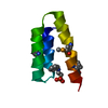

 PDBj
PDBj


 Assembly
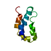

 Sample preparation
 Processing
 X-PLOR