#191 - Nov 2015 Glutamate-gated Chloride Receptors similarity (1)
-
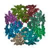
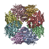
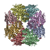
Assembly
Deposited unit
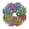
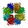
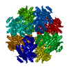
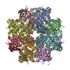
A: similar to chloromuconate cycloisomerase B: similar to chloromuconate cycloisomerase C: similar to chloromuconate cycloisomerase D: similar to chloromuconate cycloisomerase E: similar to chloromuconate cycloisomerase F: similar to chloromuconate cycloisomerase G: similar to chloromuconate cycloisomerase H: similar to chloromuconate cycloisomerase hetero molecules
Mass: 18.015 Da / Num. of mol.: 1521 / Source method: isolated from a natural source / Formula: H2O
-
Experimental details
-
Experiment
Experiment
Method: X-RAY DIFFRACTION / Number of used crystals: 1
-
Sample preparation
Crystal
Density Matthews: 2.43 Å3/Da / Density % sol: 49.48 %
Crystal grow
Temperature: 293 K / Method: vapor diffusion, hanging drop / pH: 7 Details: The protein was concentrated to 20 mg/ml and dialyzed against 5 mM HEPES, 2 mM MgCl2, 50 mM NaCl, 0.25 mM TCEP, 1 mM NaN3, pH 7.0, drop frozen as small pellets in liquid nitrogen and stored ...Details: The protein was concentrated to 20 mg/ml and dialyzed against 5 mM HEPES, 2 mM MgCl2, 50 mM NaCl, 0.25 mM TCEP, 1 mM NaN3, pH 7.0, drop frozen as small pellets in liquid nitrogen and stored at -80 C. The frozen concentrated protein was thawed and diluted to 10 mg/mL with 5 mM HEPES, 2 mM MgCl2, 1 mM NaN3, pH 7.5. A stock solution of the peptide substrate L-Ala-L-Glu (Sigma) was neutralized to pH 7.5 with NaOH and brought to a final concentration of 1.6 M. This solution was added to the protein as 1/50th part by volume and the resultant mixture utilized for crystallization experiments. The initial crystals were grown by hanging drop by mixing 5 ?l of protein solution and 5 ?l of a solution containing 9-11% dimethyl-PEG 5000, 50 mM MOPS, 1% MPD, 1 mM NaN3, pH 7.0 and suspending the droplets over 600 ?l precipitant at 20 C, VAPOR DIFFUSION, HANGING DROP, temperature 293K
In the structure databanks used in Yorodumi, some data are registered as the other names, "COVID-19 virus" and "2019-nCoV". Here are the details of the virus and the list of structure data.
Jan 31, 2019. EMDB accession codes are about to change! (news from PDBe EMDB page)
EMDB accession codes are about to change! (news from PDBe EMDB page)
The allocation of 4 digits for EMDB accession codes will soon come to an end. Whilst these codes will remain in use, new EMDB accession codes will include an additional digit and will expand incrementally as the available range of codes is exhausted. The current 4-digit format prefixed with “EMD-” (i.e. EMD-XXXX) will advance to a 5-digit format (i.e. EMD-XXXXX), and so on. It is currently estimated that the 4-digit codes will be depleted around Spring 2019, at which point the 5-digit format will come into force.
The EM Navigator/Yorodumi systems omit the EMD- prefix.
Related info.:Q: What is EMD? / ID/Accession-code notation in Yorodumi/EM Navigator
Yorodumi is a browser for structure data from EMDB, PDB, SASBDB, etc.
This page is also the successor to EM Navigator detail page, and also detail information page/front-end page for Omokage search.
The word "yorodu" (or yorozu) is an old Japanese word meaning "ten thousand". "mi" (miru) is to see.
Related info.:EMDB / PDB / SASBDB / Comparison of 3 databanks / Yorodumi Search / Aug 31, 2016. New EM Navigator & Yorodumi / Yorodumi Papers / Jmol/JSmol / Function and homology information / Changes in new EM Navigator and Yorodumi
 Movie
Movie Controller

 Yorodumi
 Open data
 Basic information
 Components
 Keywords
 Function and homology information
 X-RAY DIFFRACTION /
  Authors
 Citation
 Structure visualization
 Downloads & links
 Other downloads
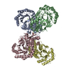


 PDBj
PDBj


 Assembly
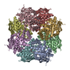

 Mass: 24.305 Da / Num. of mol.: 8 / Source method: obtained synthetically / Formula: Mg
 Type: L-peptide linking / Mass: 89.093 Da / Num. of mol.: 8 / Source method: obtained synthetically / Formula: C3H7NO2
 Type: L-peptide linking / Mass: 147.129 Da / Num. of mol.: 8 / Source method: obtained synthetically / Formula: C5H9NO4
 Mass: 18.015 Da / Num. of mol.: 1521 / Source method: isolated from a natural source / Formula: H2O
 Sample preparation
 / Beamline: 19-ID / Wavelength: 1 Å
 Processing